5A0T

Catalysis and 5' end sensing by ribonuclease RNase J of the metallo- beta-lactamase family


Experimental Data Snapshot

  • Method: X-RAY DIFFRACTION
  • Resolution: 2.28 Å
  • R-Value Free: 0.177 
  • R-Value Work: 0.145 
  • R-Value Observed: 0.146 

wwPDB Validation   3D Report Full Report


This is version 1.2 of the entry. See complete history


Literature

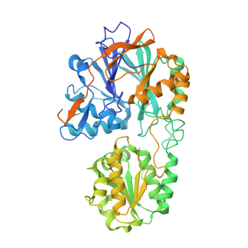
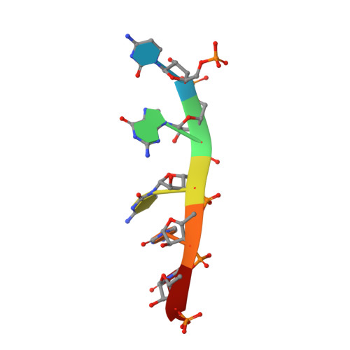
Linkage of Catalysis and 5' End Recognition in Ribonuclease Rnase J

Pei, X.Y.Bralley, P.Jones, G.H.Luisi, B.F.

(2015) Nucleic Acids Res 43: 8066

  • DOI: https://doi.org/10.1093/nar/gkv732
  • Primary Citation of Related Structures:  
    5A0T, 5A0V

  • PubMed Abstract: 

    In diverse bacterial species, the turnover and processing of many RNAs is mediated by the ribonuclease RNase J, a member of the widely occurring metallo-β-lactamase enzyme family. We present crystal structures of Streptomyces coelicolor RNase J with bound RNA in pre- and post-cleavage states, at 2.27 Å and 2.80 Å resolution, respectively. These structures reveal snapshots of the enzyme cleaving substrate directionally and sequentially from the 5' terminus. In the pre-cleavage state, a water molecule is coordinated to a zinc ion pair in the active site but is imperfectly oriented to launch a nucleophilic attack on the phosphate backbone. A conformational switch is envisaged that enables the in-line positioning of the attacking water and may be facilitated by magnesium ions. Adjacent to the scissile bond, four bases are stacked in a tightly sandwiching pocket, and mutagenesis results indicate that this organization helps to drive processive exo-ribonucleolytic cleavage. Like its numerous homologues, S. coelicolor RNase J can also cleave some RNA internally, and the structural data suggest how the preference for exo- versus endo-cleavage mode is linked with recognition of the chemical status of the substrate's 5' end.


  • Organizational Affiliation

    Department of Biochemistry, University of Cambridge, Tennis Court Road, Cambridge CB2 1GA, UK.


Macromolecules

Find similar proteins by:  (by identity cutoff)  |  3D Structure
Entity ID: 1
MoleculeChains Sequence LengthOrganismDetailsImage
RIBONUCLEASE J
A, B
561Streptomyces coelicolor A3(2)Mutation(s): 0 
UniProt
Find proteins for O86842 (Streptomyces coelicolor (strain ATCC BAA-471 / A3(2) / M145))
Explore O86842 
Go to UniProtKB:  O86842
Entity Groups  
Sequence Clusters30% Identity50% Identity70% Identity90% Identity95% Identity100% Identity
UniProt GroupO86842
Sequence Annotations
Expand
  • Reference Sequence

Find similar nucleic acids by:  Sequence   |   3D Structure  

Entity ID: 2
MoleculeChains LengthOrganismImage
5'-R(*CP*GP*CP*CP*UP)-3'C [auth E],
D [auth F]
6Escherichia coli BL21(DE3)
Sequence Annotations
Expand
  • Reference Sequence
Experimental Data & Validation

Experimental Data

  • Method: X-RAY DIFFRACTION
  • Resolution: 2.28 Å
  • R-Value Free: 0.177 
  • R-Value Work: 0.145 
  • R-Value Observed: 0.146 
  • Space Group: P 43 2 2
Unit Cell:
Length ( Å )Angle ( ˚ )
a = 186.187α = 90
b = 186.187β = 90
c = 113.382γ = 90
Software Package:
Software NamePurpose
MOSFLMdata reduction
Aimlessdata scaling
SHELXphasing
PHENIXphasing
PHENIXrefinement

Structure Validation

View Full Validation Report


Entry History 

Deposition Data

Revision History  (Full details and data files)

  • Version 1.0: 2015-08-19
    Type: Initial release
  • Version 1.1: 2015-10-07
    Changes: Database references
  • Version 1.2: 2024-01-10
    Changes: Data collection, Database references, Derived calculations, Other, Refinement description