Binding of the Lactococcal Drug Dependent Transcriptional Regulator LmrR to Its Ligands and Responsive Promoter Regions.
van der Berg, J.P., Madoori, P.K., Komarudin, A.G., Thunnissen, A.M., Driessen, A.J.(2015) PLoS One 10: e0135467-e0135467
- PubMed: 26267906 Search on PubMedSearch on PubMed Central
- DOI: https://doi.org/10.1371/journal.pone.0135467
- Primary Citation Related Structures:
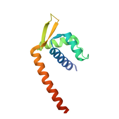
4ZZD - PubMed Abstract:
The heterodimeric ABC transporter LmrCD from Lactococcus lactis is able to extrude several different toxic compounds from the cell, fulfilling a role in the intrinsic and induced drug resistance. The expression of the lmrCD genes is regulated by the multi-drug binding repressor LmrR, which also binds to its own promoter to autoregulate its own expression. Previously, we reported the crystal structure of LmrR in the presence and absence of the drugs Hoechst 33342 and daunomycin. Analysis of the mechanism how drugs control the repressor activity of LmrR is impeded by the fact that these drugs also bind to DNA. Here we identified, using X-ray crystallography and fluorescence, that riboflavin binds into the drug binding cavity of LmrR, adopting a similar binding mode as Hoechst 33342 and daunomycin. Microscale thermophoresis was employed to quantify the binding affinity of LmrR to its responsive promoter regions and to evaluate the cognate site of LmrR in the lmrCD promoter region. Riboflavin reduces the binding affinity of LmrR for the promoter regions. Our results support a model wherein drug binding to LmrR relieves the LmrR dependent repression of the lmrCD genes.
- Molecular Microbiology, Groningen Biomolecular Sciences and Biotechnology Institute, University of Groningen, Groningen, The Netherlands.
Organizational Affiliation: