4ZXS

HSV-1 nuclear egress complex

  • Classification: VIRAL PROTEIN
  • Organism(s): Human alphaherpesvirus 1 strain 17
  • Expression System: Escherichia coli
  • Mutation(s): No 

  • Deposited: 2015-05-20 Released: 2015-11-11 
  • Deposition Author(s): Bigalke, J.M., Heldwein, E.E.
  • Funding Organization(s): German Research Foundation (DFG), National Institutes of Health/National Institute of General Medical Sciences (NIH/NIGMS), National Institutes of Health/National Institute Of Allergy and Infectious Diseases (NIH/NIAID), Burroughs Wellcome Fund

Experimental Data Snapshot

  • Method: X-RAY DIFFRACTION
  • Resolution: 2.77 Å
  • R-Value Free: 0.265 
  • R-Value Work: 0.217 
  • R-Value Observed: 0.220 

wwPDB Validation   3D Report Full Report


This is version 1.6 of the entry. See complete history


Literature

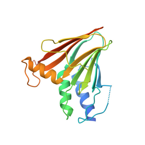
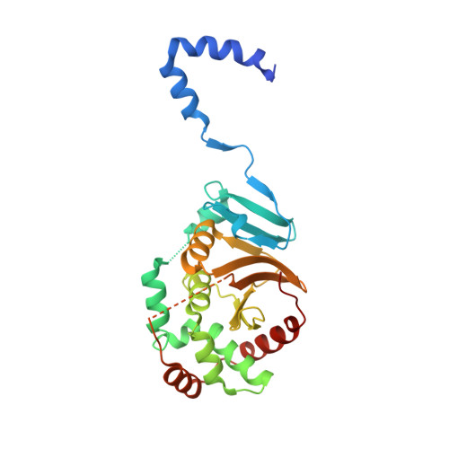
Structural basis of membrane budding by the nuclear egress complex of herpesviruses.

Bigalke, J.M.Heldwein, E.E.

(2015) EMBO J 34: 2921-2936

  • DOI: https://doi.org/10.15252/embj.201592359
  • Primary Citation of Related Structures:  
    4Z3U, 4ZXS

  • PubMed Abstract: 

    During nuclear egress, herpesvirus capsids bud at the inner nuclear membrane forming perinuclear viral particles that subsequently fuse with the outer nuclear membrane, releasing capsids into the cytoplasm. This unusual budding process is mediated by the nuclear egress complex (NEC) composed of two conserved viral proteins, UL31 and UL34. Earlier, we discovered that the herpesvirus nuclear egress complex (NEC) could bud synthetic membranes in vitro without the help of other proteins by forming a coat-like hexagonal scaffold inside the budding membrane. To understand the structural basis of NEC-mediated membrane budding, we determined the crystal structures of the NEC from two herpesviruses. The hexagonal lattice observed in the NEC crystals recapitulates the honeycomb coats within the budded vesicles. Perturbation of the oligomeric interfaces through mutagenesis blocks budding in vitro confirming that NEC oligomerization into a honeycomb lattice drives budding. The structure represents the first atomic-level view of an oligomeric array formed by a membrane-deforming protein, making possible the dissection of its unique budding mechanism and the design of inhibitors to block it.


  • Organizational Affiliation

    Department of Molecular Biology and Microbiology, Tufts University School of Medicine, Boston, MA, USA.


Macromolecules
Find similar proteins by:  (by identity cutoff)  |  3D Structure
Entity ID: 1
MoleculeChains Sequence LengthOrganismDetailsImage
Virion egress protein UL34
A, C
183Human alphaherpesvirus 1 strain 17Mutation(s): 0 
Gene Names: UL34
UniProt
Find proteins for P10218 (Human herpesvirus 1 (strain 17))
Explore P10218 
Go to UniProtKB:  P10218
Entity Groups  
Sequence Clusters30% Identity50% Identity70% Identity90% Identity95% Identity100% Identity
UniProt GroupP10218
Sequence Annotations
Expand
  • Reference Sequence
Find similar proteins by:  (by identity cutoff)  |  3D Structure
Entity ID: 2
MoleculeChains Sequence LengthOrganismDetailsImage
Virion egress protein UL31
B, D
260Human alphaherpesvirus 1 strain 17Mutation(s): 0 
Gene Names: UL31
UniProt
Find proteins for P10215 (Human herpesvirus 1 (strain 17))
Explore P10215 
Go to UniProtKB:  P10215
Entity Groups  
Sequence Clusters30% Identity50% Identity70% Identity90% Identity95% Identity100% Identity
UniProt GroupP10215
Sequence Annotations
Expand
  • Reference Sequence
Small Molecules
Ligands 4 Unique
IDChains Name / Formula / InChI Key2D Diagram3D Interactions
ZN
Query on ZN

Download Ideal Coordinates CCD File 
H [auth B],
L [auth D]
ZINC ION
Zn
PTFCDOFLOPIGGS-UHFFFAOYSA-N
NI
Query on NI

Download Ideal Coordinates CCD File 
E [auth A],
J [auth C]
NICKEL (II) ION
Ni
VEQPNABPJHWNSG-UHFFFAOYSA-N
CL
Query on CL

Download Ideal Coordinates CCD File 
F [auth A],
G [auth A],
K [auth C]
CHLORIDE ION
Cl
VEXZGXHMUGYJMC-UHFFFAOYSA-M
NA
Query on NA

Download Ideal Coordinates CCD File 
I [auth B],
M [auth D]
SODIUM ION
Na
FKNQFGJONOIPTF-UHFFFAOYSA-N
Experimental Data & Validation

Experimental Data

Unit Cell:
Length ( Å )Angle ( ˚ )
a = 110.529α = 90
b = 110.529β = 90
c = 155.85γ = 120
Software Package:
Software NamePurpose
PHENIXrefinement
Cootmodel building
PHASERphasing
XDSdata scaling
XDSdata reduction

Structure Validation

View Full Validation Report


Entry History & Funding Information

Deposition Data


Funding OrganizationLocationGrant Number
German Research Foundation (DFG)GermanyBI 1658/1-1
National Institutes of Health/National Institute of General Medical Sciences (NIH/NIGMS)United States1R01GM111795-01
National Institutes of Health/National Institute Of Allergy and Infectious Diseases (NIH/NIAID)United States1R21AI097573-01A1
Burroughs Wellcome FundUnited States--

Revision History  (Full details and data files)

  • Version 1.0: 2015-11-11
    Type: Initial release
  • Version 1.1: 2015-12-16
    Changes: Database references
  • Version 1.2: 2017-09-20
    Changes: Author supporting evidence, Database references, Derived calculations, Refinement description
  • Version 1.3: 2017-09-27
    Changes: Author supporting evidence
  • Version 1.4: 2018-01-17
    Changes: Author supporting evidence
  • Version 1.5: 2019-12-11
    Changes: Author supporting evidence
  • Version 1.6: 2023-09-27
    Changes: Data collection, Database references, Derived calculations, Refinement description