Activity Augmentation of Amphioxus Peptidoglycan Recognition Protein BbtPGRP3 via Fusion with a Chitin Binding Domain
Wang, W.J., Cheng, W., Luo, M., Yan, Q., Yu, H.M., Li, Q., Cao, D.D., Huang, S., Xu, A., Mariuzza, R.A., Chen, Y., Zhou, C.Z.(2015) PLoS One 10: e0140953-e0140953
- PubMed: 26479246 Search on PubMedSearch on PubMed Central
- DOI: https://doi.org/10.1371/journal.pone.0140953
- Primary Citation Related Structures:
4Z8I, 4ZXM - PubMed Abstract:
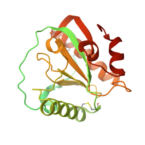
Peptidoglycan recognition proteins (PGRPs), which have been identified in most animals, are pattern recognition molecules that involve antimicrobial defense. Resulting from extraordinary expansion of innate immune genes, the amphioxus encodes many PGRPs of diverse functions. For instance, three isoforms of PGRP encoded by Branchiostoma belcheri tsingtauense, termed BbtPGRP1~3, are fused with a chitin binding domain (CBD) at the N-terminus. Here we report the 2.7 Å crystal structure of BbtPGRP3, revealing an overall structure of an N-terminal hevein-like CBD followed by a catalytic PGRP domain. Activity assays combined with site-directed mutagenesis indicated that the individual PGRP domain exhibits amidase activity towards both DAP-type and Lys-type peptidoglycans (PGNs), the former of which is favored. The N-terminal CBD not only has the chitin-binding activity, but also enables BbtPGRP3 to gain a five-fold increase of amidase activity towards the Lys-type PGNs, leading to a significantly broadened substrate spectrum. Together, we propose that modular evolution via domain shuffling combined with gene horizontal transfer makes BbtPGRP1~3 novel PGRPs of augmented catalytic activity and broad recognition spectrum.
- Hefei National Laboratory for Physical Sciences at Microscale and the Innovation Center for Cell Signaling Network, School of Life Sciences, University of Science and Technology of China, Hefei, Anhui, China.
Organizational Affiliation: