4ZW2

Crystal structure of the Mouse voltage gated calcium channel beta subunit isoform 1a in complex with Alpha Interaction Domain peptide.


Experimental Data Snapshot

  • Method: X-RAY DIFFRACTION
  • Resolution: 1.86 Å
  • R-Value Free: 0.207 
  • R-Value Work: 0.169 
  • R-Value Observed: 0.171 

wwPDB Validation   3D Report Full Report


This is version 1.3 of the entry. See complete history


Literature

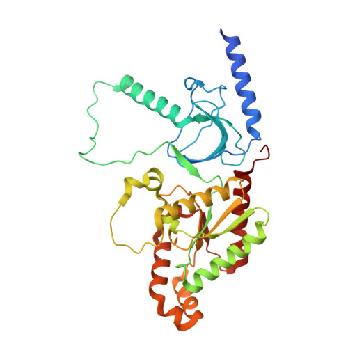
Structural and biophysical analyses of the skeletal dihydropyridine receptor beta subunit beta 1a reveal critical roles of domain interactions for stability.

Norris, N.C.Joseph, S.Aditya, S.Karunasekara, Y.Board, P.G.Dulhunty, A.F.Oakley, A.J.Casarotto, M.G.

(2017) J Biol Chem 292: 8401-8411

  • DOI: https://doi.org/10.1074/jbc.M116.763896
  • Primary Citation of Related Structures:  
    4ZW2

  • PubMed Abstract: 

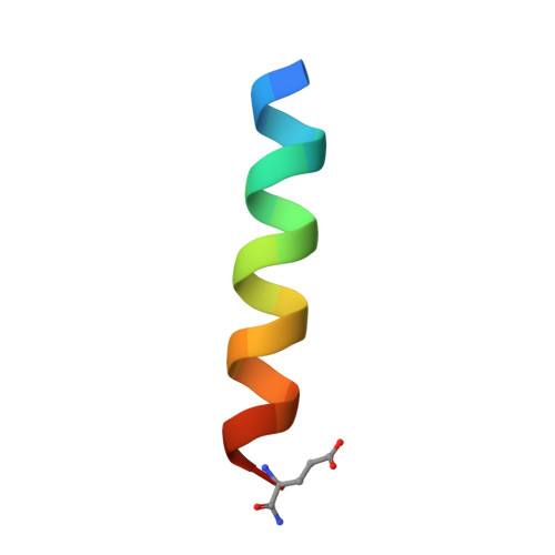
    Excitation-contraction (EC) coupling in skeletal muscle requires a physical interaction between the voltage-gated calcium channel dihydropyridine receptor (DHPR) and the ryanodine receptor Ca 2+ release channel. Although the exact molecular mechanism that initiates skeletal EC coupling is unresolved, it is clear that both the α 1 and β subunits of DHPR are essential for this process. Here, we employed a series of techniques, including size-exclusion chromatography-multi-angle light scattering, differential scanning fluorimetry, and isothermal calorimetry, to characterize various biophysical properties of the skeletal DHPR β subunit β 1a Removal of the intrinsically disordered N and C termini and the hook region of β 1a prevented oligomerization, allowing for its structural determination by X-ray crystallography. The structure had a topology similar to that of previously determined β isoforms, which consist of SH3 and guanylate kinase domains. However, transition melting temperatures derived from the differential scanning fluorimetry experiments indicated a significant difference in stability of ∼2-3 °C between the β 1a and β 2a constructs, and the addition of the DHPR α 1s I-II loop (α-interaction domain) peptide stabilized both β isoforms by ∼6-8 °C. Similar to other β isoforms, β 1a bound with nanomolar affinity to the α-interaction domain, but binding affinities were influenced by amino acid substitutions in the adjacent SH3 domain. These results suggest that intramolecular interactions between the SH3 and guanylate kinase domains play a role in the stability of β 1a while also providing a conduit for allosteric signaling events.


  • Organizational Affiliation

    John Curtin School of Medical Research, Australian National University, Canberra, Australian Capital Territory 2601, Australia.


Macromolecules
Find similar proteins by:  (by identity cutoff)  |  3D Structure
Entity ID: 1
MoleculeChains Sequence LengthOrganismDetailsImage
Voltage-dependent L-type calcium channel subunit beta-1,Voltage-dependent L-type calcium channel subunit beta-1333Mus musculusMutation(s): 0 
Gene Names: Cacnb1Cacnlb1
UniProt
Find proteins for Q8R3Z5 (Mus musculus)
Explore Q8R3Z5 
Go to UniProtKB:  Q8R3Z5
Entity Groups  
Sequence Clusters30% Identity50% Identity70% Identity90% Identity95% Identity100% Identity
UniProt GroupQ8R3Z5
Sequence Annotations
Expand
  • Reference Sequence

Find similar proteins by:  Sequence   |   3D Structure  

Entity ID: 2
MoleculeChains Sequence LengthOrganismDetailsImage
Voltage-dependent L-type calcium channel subunit alpha-1S20Mus musculusMutation(s): 2 
UniProt & NIH Common Fund Data Resources
Find proteins for Q02789 (Mus musculus)
Explore Q02789 
Go to UniProtKB:  Q02789
IMPC:  MGI:88294
Entity Groups  
Sequence Clusters30% Identity50% Identity70% Identity90% Identity95% Identity100% Identity
UniProt GroupQ02789
Sequence Annotations
Expand
  • Reference Sequence
Small Molecules
Experimental Data & Validation

Experimental Data

  • Method: X-RAY DIFFRACTION
  • Resolution: 1.86 Å
  • R-Value Free: 0.207 
  • R-Value Work: 0.169 
  • R-Value Observed: 0.171 
  • Space Group: P 21 21 21
Unit Cell:
Length ( Å )Angle ( ˚ )
a = 46.24α = 90
b = 69.371β = 90
c = 131.104γ = 90
Software Package:
Software NamePurpose
REFMACrefinement
DENZOdata reduction
SCALEPACKdata scaling
PHASERphasing

Structure Validation

View Full Validation Report


Entry History 

Deposition Data

Revision History  (Full details and data files)

  • Version 1.0: 2016-06-01
    Type: Initial release
  • Version 1.1: 2016-07-27
    Changes: Data collection
  • Version 1.2: 2017-08-02
    Changes: Database references, Derived calculations
  • Version 1.3: 2023-09-27
    Changes: Data collection, Database references, Refinement description