4ZUU | pdb_00004zuu

Crystal structure of Equine MHC I(Eqca-N*00602) in complexed with equine infectious anaemia virus (EIAV) derived peptide Gag-CF9


Experimental Data Snapshot

  • Method: X-RAY DIFFRACTION
  • Resolution: 2.20 Å
  • R-Value Free: 
    0.266 (Depositor), 0.266 (DCC) 
  • R-Value Work: 
    0.228 (Depositor), 0.230 (DCC) 
  • R-Value Observed: 
    0.230 (Depositor) 

Starting Model: experimental
View more details

wwPDB Validation 3D Report Full Report

Validation slider image for 4ZUU

This is version 1.2 of the entry. See complete history

Literature

Structural Illumination of Equine MHC Class I Molecules Highlights Unconventional Epitope Presentation Manner That Is Evolved in Equine Leukocyte Antigen Alleles

Yao, S.Liu, J.Qi, J.Chen, R.Zhang, N.Liu, Y.Wang, J.Wu, Y.Gao, G.F.Xia, C.

(2016) J Immunol 196: 1943-1954

  • DOI: https://doi.org/10.4049/jimmunol.1501352
  • Primary Citation Related Structures: 
    4ZUS, 4ZUT, 4ZUU, 4ZUV, 4ZUW

  • PubMed Abstract: 

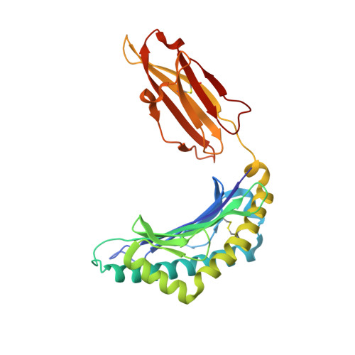
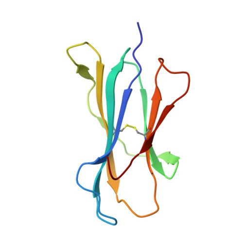
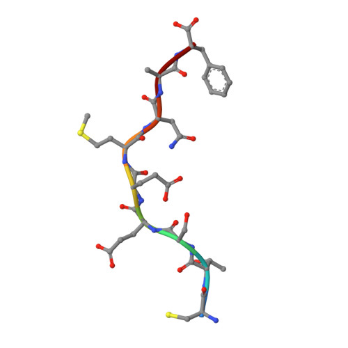
    MHC class I (MHC I)-restricted virus-specific CTLs are implicated as critical components in the control of this naturally occurring lentivirus and in the protective immune response to the successfully applied attenuated equine infectious anemia virus vaccine in the horse. Nevertheless, the structural basis for how the equine MHC I presents epitope peptides remains unknown. In this study, we investigated the binding of several equine infectious anemia virus-derived epitope peptides by the ability to refold recombinant molecules and by thermal stability, and then by determining the x-ray structure of five peptide-MHC I complexes: equine MHC class I allele (Eqca)-N*00602/Env-RW12, Eqca-N*00602/Gag-GW12, Eqca-N*00602/Rev-QW11, Eqca-N*00602/Gag-CF9, and Eqca-N*00601/Gag-GW12. Although Eqca-N*00601 and Eqca-N*00602 differ by a single amino acid, Eqca-N*00601 exhibited a drastically different peptide presentation when binding a similar CTL epitope, Gag-GW12; the result makes the previously reported function clear to be non-cross-recognition between these two alleles. The structures plus Eqca-N*00602 complexed with a 9-mer peptide are particularly noteworthy in that we illuminated differences in apparent flexibility in the center of the epitope peptides for the complexes with Gag-GW12 as compared with Env-RW12, and a strict selection of epitope peptides with normal length. The featured preferences and unconventional presentations of long peptides by equine MHC I molecules provide structural bases to explain the exceptional anti-lentivirus immunity in the horse. We think that the beneficial reference points could serve as an initial platform for other human or animal lentiviruses.


  • Organizational Affiliation
    • Department of Microbiology and Immunology, College of Veterinary Medicine, China Agricultural University, Beijing 100193, China; Department of Medical Microbiology and Immunology, University of Alberta, Edmonton, Alberta T6G 2R3, Canada;

Macromolecule Content 

  • Total Structure Weight: 44.34 kDa 
  • Atom Count: 3,246 
  • Modeled Residue Count: 382 
  • Deposited Residue Count: 382 
  • Unique protein chains: 3

Macromolecules

Find similar proteins by:|  3D Structure
Entity ID: 1
MoleculeChains  Sequence LengthOrganismDetailsImage
Classical MHC class I antigen274Equus caballusMutation(s): 0 
UniProt
Find proteins for Q860N6 (Equus caballus)
Explore Q860N6 
Go to UniProtKB:  Q860N6
Entity Groups
Sequence Clusters30% Identity50% Identity70% Identity90% Identity95% Identity100% Identity
UniProt GroupQ860N6
Sequence Annotations
Expand
Reference Sequence
Find similar proteins by:|  3D Structure
Entity ID: 2
MoleculeChains  Sequence LengthOrganismDetailsImage
Beta-2-microglobulin99Mus musculusMutation(s): 1 
Gene Names: B2m
UniProt & NIH Common Fund Data Resources
Find proteins for P01887 (Mus musculus)
Explore P01887 
Go to UniProtKB:  P01887
IMPC:  MGI:88127
Entity Groups
Sequence Clusters30% Identity50% Identity70% Identity90% Identity95% Identity100% Identity
UniProt GroupP01887
Sequence Annotations
Expand
Reference Sequence
Find similar proteins by:  Sequence   |   3D Structure  
Entity ID: 3
MoleculeChains  Sequence LengthOrganismDetailsImage
CYS-THR-SER-GLU-GLU-MET-ASN-ALA-PHE9Equine infectious anemia virusMutation(s): 0 
UniProt
Find proteins for P69732 (Equine infectious anemia virus (strain Wyoming))
Explore P69732 
Go to UniProtKB:  P69732
Entity Groups
UniProt GroupP69732
Sequence Annotations
Expand
Reference Sequence

Experimental Data & Validation

Experimental Data

  • Method: X-RAY DIFFRACTION
  • Resolution: 2.20 Å
  • R-Value Free:  0.266 (Depositor), 0.266 (DCC) 
  • R-Value Work:  0.228 (Depositor), 0.230 (DCC) 
  • R-Value Observed: 0.230 (Depositor) 
Space Group: P 41 21 2
Unit Cell:
Length ( Å )Angle ( ˚ )
a = 69.47α = 90
b = 69.47β = 90
c = 206.458γ = 90
Software Package:
Software NamePurpose
PHENIXrefinement
HKL-2000data reduction
HKL-2000data scaling
HKL-2000phasing

Structure Validation

View Full Validation Report


Entry History 

Deposition Data

Revision History  (Full details and data files)

  • Version 1.0: 2016-04-06
    Type: Initial release
  • Version 1.1: 2023-11-08
    Changes: Data collection, Database references, Derived calculations, Refinement description
  • Version 1.2: 2024-10-30
    Changes: Structure summary