4ZR1

Hydroxylase domain of scs7p


Experimental Data Snapshot

  • Method: X-RAY DIFFRACTION
  • Resolution: 2.60 Å
  • R-Value Free: 0.239 
  • R-Value Work: 0.197 
  • R-Value Observed: 0.199 

wwPDB Validation   3D Report Full Report


Ligand Structure Quality Assessment 


This is version 1.6 of the entry. See complete history


Literature

The Crystal Structure of an Integral Membrane Fatty Acid alpha-Hydroxylase.

Zhu, G.Koszelak-Rosenblum, M.Connelly, S.M.Dumont, M.E.Malkowski, M.G.

(2015) J Biol Chem 290: 29820-29833

  • DOI: https://doi.org/10.1074/jbc.M115.680124
  • Primary Citation of Related Structures:  
    4ZR0, 4ZR1

  • PubMed Abstract: 

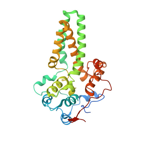
    Neuronal electrical impulse propagation is facilitated by the myelin sheath, a compact membrane surrounding the axon. The myelin sheath is highly enriched in galactosylceramide (GalCer) and its sulfated derivative sulfatide. Over 50% of GalCer and sulfatide in myelin is hydroxylated by the integral membrane enzyme fatty acid 2-hydroxylase (FA2H). GalCer hydroxylation contributes to the compact nature of the myelin membrane, and mutations in FA2H result in debilitating leukodystrophies and spastic paraparesis. We report here the 2.6 Å crystal structure of sphingolipid α-hydroxylase (Scs7p), a yeast homolog of FA2H. The Scs7p core is composed of a helical catalytic cap domain that sits atop four transmembrane helices that anchor the enzyme in the endoplasmic reticulum. The structure contains two zinc atoms coordinated by the side chains of 10 highly conserved histidines within a dimetal center located near the plane of the cytosolic membrane. We used a yeast genetic approach to confirm the important role of the dimetal-binding histidines in catalysis and identified Tyr-322 and Asp-323 as critical determinants involved in the hydroxylase reaction. Examination of the Scs7p structure, coupled with molecular dynamics simulations, allowed for the generation of a model of ceramide binding to Scs7p. Comparison of the Scs7p structure and substrate-binding model to the structure of steroyl-CoA desaturase revealed significant differences in the architecture of the catalytic cap domain and location of the dimetal centers with respect to the membrane. These observations provide insight into the different mechanisms of substrate binding and recognition of substrates by the hydroxylase and desaturase enzymes.


  • Organizational Affiliation

    From the Hauptman-Woodward Medical Research Institute, Buffalo, New York 14203.


Macromolecules
Find similar proteins by:  (by identity cutoff)  |  3D Structure
Entity ID: 1
MoleculeChains Sequence LengthOrganismDetailsImage
Ceramide very long chain fatty acid hydroxylase SCS7
A, B
298Saccharomyces cerevisiae S288CMutation(s): 0 
Gene Names: SCS7FAH1YMR272CYM8156.14C
EC: 1
Membrane Entity: Yes 
UniProt
Find proteins for Q03529 (Saccharomyces cerevisiae (strain ATCC 204508 / S288c))
Explore Q03529 
Go to UniProtKB:  Q03529
Entity Groups  
Sequence Clusters30% Identity50% Identity70% Identity90% Identity95% Identity100% Identity
UniProt GroupQ03529
Sequence Annotations
Expand
  • Reference Sequence
Small Molecules
Ligands 3 Unique
IDChains Name / Formula / InChI Key2D Diagram3D Interactions
BOG
Query on BOG

Download Ideal Coordinates CCD File 
E [auth A]
F [auth A]
G [auth A]
H [auth A]
I [auth A]
E [auth A],
F [auth A],
G [auth A],
H [auth A],
I [auth A],
J [auth A],
O [auth B],
P [auth B]
octyl beta-D-glucopyranoside
C14 H28 O6
HEGSGKPQLMEBJL-RKQHYHRCSA-N
TRD
Query on TRD

Download Ideal Coordinates CCD File 
K [auth A]
L [auth A]
Q [auth B]
R [auth B]
S [auth B]
K [auth A],
L [auth A],
Q [auth B],
R [auth B],
S [auth B],
T [auth B],
U [auth B],
V [auth B]
TRIDECANE
C13 H28
IIYFAKIEWZDVMP-UHFFFAOYSA-N
ZN
Query on ZN

Download Ideal Coordinates CCD File 
C [auth A],
D [auth A],
M [auth B],
N [auth B]
ZINC ION
Zn
PTFCDOFLOPIGGS-UHFFFAOYSA-N
Experimental Data & Validation

Experimental Data

  • Method: X-RAY DIFFRACTION
  • Resolution: 2.60 Å
  • R-Value Free: 0.239 
  • R-Value Work: 0.197 
  • R-Value Observed: 0.199 
  • Space Group: I 21 3
Unit Cell:
Length ( Å )Angle ( ˚ )
a = 202.762α = 90
b = 202.762β = 90
c = 202.762γ = 90
Software Package:
Software NamePurpose
PHENIXrefinement
HKL-2000data reduction
HKL-2000data scaling
PHENIXphasing

Structure Validation

View Full Validation Report


Ligand Structure Quality Assessment 


Entry History & Funding Information

Deposition Data


Funding OrganizationLocationGrant Number
National Institutes of Health/National Institute of General Medical Sciences (NIH/NIGMS)United StatesU54 GM094611

Revision History  (Full details and data files)

  • Version 1.0: 2015-07-29
    Type: Initial release
  • Version 1.1: 2015-11-18
    Changes: Database references
  • Version 1.2: 2015-12-23
    Changes: Database references
  • Version 1.3: 2017-09-06
    Changes: Author supporting evidence, Database references, Derived calculations
  • Version 1.4: 2019-12-25
    Changes: Author supporting evidence
  • Version 1.5: 2020-07-29
    Type: Remediation
    Reason: Carbohydrate remediation
    Changes: Data collection, Derived calculations, Structure summary
  • Version 1.6: 2024-03-06
    Changes: Data collection, Database references, Structure summary