4ZNO

Crystal structure of Dln1 complexed with sucrose


Experimental Data Snapshot

  • Method: X-RAY DIFFRACTION
  • Resolution: 1.86 Å
  • R-Value Free: 0.225 
  • R-Value Work: 0.189 
  • R-Value Observed: 0.191 

wwPDB Validation   3D Report Full Report


This is version 2.1 of the entry. See complete history


Literature

Structural basis for receptor recognition and pore formation of a zebrafish aerolysin-like protein.

Jia, N.Liu, N.Cheng, W.Jiang, Y.L.Sun, H.Chen, L.L.Peng, J.Zhang, Y.Ding, Y.H.Zhang, Z.H.Wang, X.Cai, G.Wang, J.Dong, M.Q.Zhang, Z.Wu, H.Wang, H.W.Chen, Y.Zhou, C.Z.

(2016) EMBO Rep 17: 235-248

  • DOI: https://doi.org/10.15252/embr.201540851
  • Primary Citation of Related Structures:  
    4ZNO, 4ZNQ, 4ZNR, 5DI0

  • PubMed Abstract: 

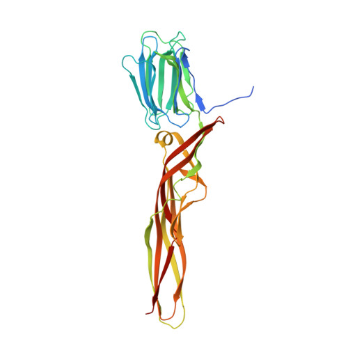
    Various aerolysin-like pore-forming proteins have been identified from bacteria to vertebrates. However, the mechanism of receptor recognition and/or pore formation of the eukaryotic members remains unknown. Here, we present the first crystal and electron microscopy structures of a vertebrate aerolysin-like protein from Danio rerio, termed Dln1, before and after pore formation. Each subunit of Dln1 dimer comprises a β-prism lectin module followed by an aerolysin module. Specific binding of the lectin module toward high-mannose glycans triggers drastic conformational changes of the aerolysin module in a pH-dependent manner, ultimately resulting in the formation of a membrane-bound octameric pore. Structural analyses combined with computational simulations and biochemical assays suggest a pore-forming process with an activation mechanism distinct from the previously characterized bacterial members. Moreover, Dln1 and its homologs are ubiquitously distributed in bony fishes and lamprey, suggesting a novel fish-specific defense molecule.


  • Organizational Affiliation

    Hefei National Laboratory for Physical Sciences at the Microscale and School of Life Sciences, University of Science and Technology of China, Hefei, China.


Macromolecules
Find similar proteins by:  (by identity cutoff)  |  3D Structure
Entity ID: 1
MoleculeChains Sequence LengthOrganismDetailsImage
Natterin-like protein
A, B
335Danio rerioMutation(s): 0 
Gene Names: zgc:113413
UniProt
Find proteins for Q5CZR5 (Danio rerio)
Explore Q5CZR5 
Go to UniProtKB:  Q5CZR5
Entity Groups  
Sequence Clusters30% Identity50% Identity70% Identity90% Identity95% Identity100% Identity
UniProt GroupQ5CZR5
Sequence Annotations
Expand
  • Reference Sequence
Oligosaccharides

Help

Entity ID: 2
MoleculeChains Length2D Diagram Glycosylation3D Interactions
beta-D-fructofuranose-(2-1)-alpha-D-glucopyranose
C, D
2N/A
Glycosylation Resources
GlyTouCan:  G05551OP
GlyCosmos:  G05551OP
Biologically Interesting Molecules (External Reference) 1 Unique
Experimental Data & Validation

Experimental Data

  • Method: X-RAY DIFFRACTION
  • Resolution: 1.86 Å
  • R-Value Free: 0.225 
  • R-Value Work: 0.189 
  • R-Value Observed: 0.191 
  • Space Group: P 21 21 21
Unit Cell:
Length ( Å )Angle ( ˚ )
a = 57.642α = 90
b = 95.169β = 90
c = 148.921γ = 90
Software Package:
Software NamePurpose
REFMACrefinement
HKL-2000data reduction
HKL-3000data scaling
MOLREPphasing

Structure Validation

View Full Validation Report


Entry History & Funding Information

Deposition Data


Funding OrganizationLocationGrant Number
the Ministry of Science and Technology of ChinaChina2013CB835300
the Ministry of Science and Technology of ChinaChina2015CB910100
the Ministry of Science and Technology of ChinaChina2014CB910100

Revision History  (Full details and data files)

  • Version 1.0: 2016-01-20
    Type: Initial release
  • Version 1.1: 2016-03-30
    Changes: Database references
  • Version 2.0: 2020-07-29
    Type: Remediation
    Reason: Carbohydrate remediation
    Changes: Atomic model, Data collection, Derived calculations, Non-polymer description, Structure summary
  • Version 2.1: 2024-03-20
    Changes: Data collection, Database references, Structure summary