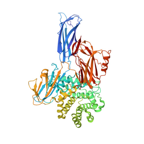
4ZIU

Crystal structure of native alpha-2-macroglobulin from Escherichia coli spanning the residues from domain MG7 to the C-terminus.


Experimental Data Snapshot

  • Method: X-RAY DIFFRACTION
  • Resolution: 2.70 Å
  • R-Value Free: 0.240 
  • R-Value Work: 0.190 
  • R-Value Observed: 0.192 

wwPDB Validation   3D Report Full Report


This is version 1.3 of the entry. See complete history


Literature

Structural and functional insights into Escherichia coli alpha 2-macroglobulin endopeptidase snap-trap inhibition.

Garcia-Ferrer, I.Arede, P.Gomez-Blanco, J.Luque, D.Duquerroy, S.Caston, J.R.Goulas, T.Gomis-Ruth, F.X.

(2015) Proc Natl Acad Sci U S A 112: 8290-8295

  • DOI: https://doi.org/10.1073/pnas.1506538112
  • Primary Citation of Related Structures:  
    4ZIQ, 4ZIU, 4ZJG, 4ZJH, 5A42

  • PubMed Abstract: 

    The survival of commensal bacteria requires them to evade host peptidases. Gram-negative bacteria from the human gut microbiome encode a relative of the human endopeptidase inhibitor, α2-macroglobulin (α2M). Escherichia coli α2M (ECAM) is a ∼ 180-kDa multidomain membrane-anchored pan-peptidase inhibitor, which is cleaved by host endopeptidases in an accessible bait region. Structural studies by electron microscopy and crystallography reveal that this cleavage causes major structural rearrangement of more than half the 13-domain structure from a native to a compact induced form. It also exposes a reactive thioester bond, which covalently traps the peptidase. Subsequently, peptidase-laden ECAM is shed from the membrane and may dimerize. Trapped peptidases are still active except against very large substrates, so inhibition potentially prevents damage of large cell envelope components, but not host digestion. Mechanistically, these results document a novel monomeric "snap trap."


  • Organizational Affiliation

    Proteolysis Lab, Department of Structural Biology ("María de Maeztu" Unit of Excellence), Molecular Biology Institute of Barcelona, Spanish Research Council (CSIC), 08028 Barcelona, Spain;


Macromolecules
Find similar proteins by:  (by identity cutoff)  |  3D Structure
Entity ID: 1
MoleculeChains Sequence LengthOrganismDetailsImage
Uncharacterized lipoprotein YfhM639Escherichia coli K-12Mutation(s): 0 
Gene Names: yfhMb2520JW2504
UniProt
Find proteins for P76578 (Escherichia coli (strain K12))
Explore P76578 
Go to UniProtKB:  P76578
Entity Groups  
Sequence Clusters30% Identity50% Identity70% Identity90% Identity95% Identity100% Identity
UniProt GroupP76578
Sequence Annotations
Expand
  • Reference Sequence
Experimental Data & Validation

Experimental Data

  • Method: X-RAY DIFFRACTION
  • Resolution: 2.70 Å
  • R-Value Free: 0.240 
  • R-Value Work: 0.190 
  • R-Value Observed: 0.192 
  • Space Group: I 2 2 2
Unit Cell:
Length ( Å )Angle ( ˚ )
a = 86.72α = 90
b = 136.19β = 90
c = 172.8γ = 90
Software Package:
Software NamePurpose
XDSdata reduction
XSCALEdata scaling
SHELXDEphasing
Cootmodel building
BUSTERrefinement

Structure Validation

View Full Validation Report


Entry History & Funding Information

Deposition Data


Funding OrganizationLocationGrant Number
European CommunitySpainFP7-PEOPLE-2011-ITN-290246
European CommunitySpainFP7-HEALTH-2012-306029-2
Spanish Ministry for Education, Culture and SportSpainAP2010-3799

Revision History  (Full details and data files)

  • Version 1.0: 2015-06-10
    Type: Initial release
  • Version 1.1: 2015-07-08
    Changes: Database references
  • Version 1.2: 2015-07-15
    Changes: Database references
  • Version 1.3: 2019-02-20
    Changes: Advisory, Data collection, Derived calculations