Regulation of endoplasmic reticulum turnover by selective autophagy.
Khaminets, A., Heinrich, T., Mari, M., Grumati, P., Huebner, A.K., Akutsu, M., Liebmann, L., Stolz, A., Nietzsche, S., Koch, N., Mauthe, M., Katona, I., Qualmann, B., Weis, J., Reggiori, F., Kurth, I., Hubner, C.A., Dikic, I.(2015) Nature 522: 354-358
- PubMed: 26040720 Search on PubMed
- DOI: https://doi.org/10.1038/nature14498
- Primary Citation Related Structures:
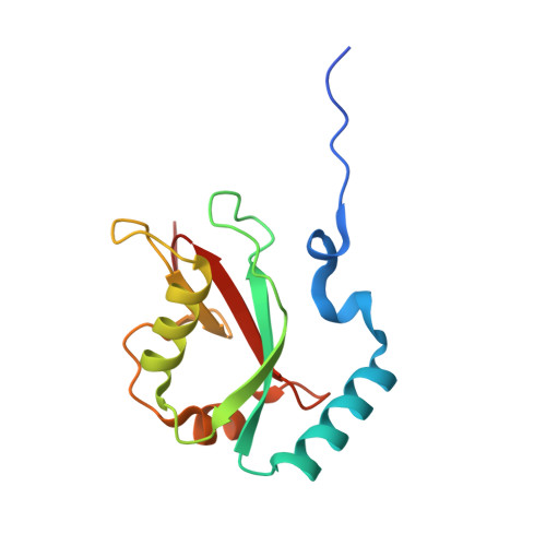
4ZDV - PubMed Abstract:
The endoplasmic reticulum (ER) is the largest intracellular endomembrane system, enabling protein and lipid synthesis, ion homeostasis, quality control of newly synthesized proteins and organelle communication. Constant ER turnover and modulation is needed to meet different cellular requirements and autophagy has an important role in this process. However, its underlying regulatory mechanisms remain unexplained. Here we show that members of the FAM134 reticulon protein family are ER-resident receptors that bind to autophagy modifiers LC3 and GABARAP, and facilitate ER degradation by autophagy ('ER-phagy'). Downregulation of FAM134B protein in human cells causes an expansion of the ER, while FAM134B overexpression results in ER fragmentation and lysosomal degradation. Mutant FAM134B proteins that cause sensory neuropathy in humans are unable to act as ER-phagy receptors. Consistently, disruption of Fam134b in mice causes expansion of the ER, inhibits ER turnover, sensitizes cells to stress-induced apoptotic cell death and leads to degeneration of sensory neurons. Therefore, selective ER-phagy via FAM134 proteins is indispensable for mammalian cell homeostasis and controls ER morphology and turnover in mice and humans.
- Institute of Biochemistry II, Goethe University School of Medicine, Theodor-Stern-Kai 7, 60590 Frankfurt am Main, Germany.
Organizational Affiliation: