


crystal structure of isocitrate dehydrogenase in complex with isocitrate and Mn from M. smegmatis
Pojer, F., Murima, P., McKinney, J.D.To be published.
Experimental Data Snapshot
Entity ID: 1 | |||||
|---|---|---|---|---|---|
| Molecule | Chains | Sequence Length | Organism | Details | Image |
| Isocitrate dehydrogenase (NADP) Icd2 | 767 | Mycolicibacterium smegmatis | Mutation(s): 0 Gene Names: icd2, MSMEG_1654, MSMEI_1615 EC: 1.1.1.42 | 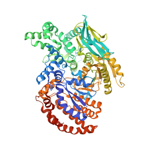 | |
UniProt | |||||
Entity Groups | |||||
| Sequence Clusters | 30% Identity50% Identity70% Identity90% Identity95% Identity100% Identity | ||||
| UniProt Group | A0QSZ3 | ||||
Sequence AnnotationsExpand | |||||
Reference Sequence | |||||
| Ligands 2 Unique | |||||
|---|---|---|---|---|---|
| ID | Chains | Name / Formula / InChI Key | 2D Diagram | 3D Interactions | |
| ICT Download:Ideal Coordinates CCD File | H [auth A] J [auth B] L [auth C] N [auth D] P [auth E] | ISOCITRIC ACID C6 H8 O7 ODBLHEXUDAPZAU-ZAFYKAAXSA-N |  | ||
| MN Download:Ideal Coordinates CCD File | G [auth A] I [auth B] K [auth C] M [auth D] O [auth E] | MANGANESE (II) ION Mn WAEMQWOKJMHJLA-UHFFFAOYSA-N |  | ||
| Length ( Å ) | Angle ( ˚ ) |
|---|---|
| a = 201.774 | α = 90 |
| b = 206.162 | β = 90.97 |
| c = 145.598 | γ = 90 |
| Software Name | Purpose |
|---|---|
| REFMAC | refinement |
| XDS | data reduction |
| XDS | data scaling |
| PHASER | phasing |


