4ZCF

Structural basis of asymmetric DNA methylation and ATP-triggered long-range diffusion by EcoP15I


Experimental Data Snapshot

  • Method: X-RAY DIFFRACTION
  • Resolution: 2.60 Å
  • R-Value Free: 0.263 
  • R-Value Work: 0.218 
  • R-Value Observed: 0.221 

wwPDB Validation   3D Report Full Report


Ligand Structure Quality Assessment 


This is version 2.0 of the entry. See complete history


Literature

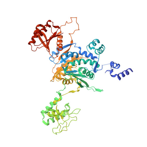
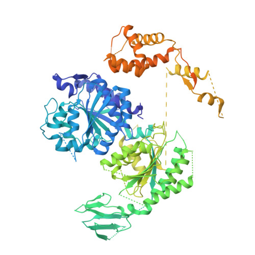
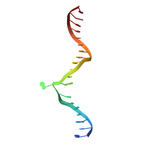
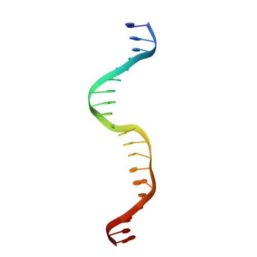
Structural basis of asymmetric DNA methylation and ATP-triggered long-range diffusion by EcoP15I.

Gupta, Y.K.Chan, S.H.Xu, S.Y.Aggarwal, A.K.

(2015) Nat Commun 6: 7363-7363

  • DOI: 10.1038/ncomms8363
  • Primary Citation of Related Structures:  
    4ZCF

  • PubMed Abstract: 
  • Type III R-M enzymes were identified >40 years ago and yet there is no structural information on these multisubunit enzymes. Here we report the structure of a Type III R-M system, consisting of the entire EcoP15I complex (Mod2Res1) bound to DNA. The structure suggests how ATP hydrolysis is coupled to long-range diffusion of a helicase on DNA, and how a dimeric methyltransferase functions to methylate only one of the two DNA strands ...

    Type III R-M enzymes were identified >40 years ago and yet there is no structural information on these multisubunit enzymes. Here we report the structure of a Type III R-M system, consisting of the entire EcoP15I complex (Mod2Res1) bound to DNA. The structure suggests how ATP hydrolysis is coupled to long-range diffusion of a helicase on DNA, and how a dimeric methyltransferase functions to methylate only one of the two DNA strands. We show that the EcoP15I motor domains are specifically adapted to bind double-stranded DNA and to facilitate DNA sliding via a novel 'Pin' domain. We also uncover unexpected 'division of labour', where one Mod subunit recognizes DNA, while the other Mod subunit methylates the target adenine--a mechanism that may extend to adenine N6 RNA methylation in mammalian cells. Together the structure sheds new light on the mechanisms of both helicases and methyltransferases in DNA and RNA metabolism.


    Organizational Affiliation

    Department of Structural and Chemical Biology, Icahn School of Medicine at Mount Sinai, Box 1677, 1425 Madison Avenue, New York, New York 10029, USA.


Macromolecules

Find similar proteins by:  (by identity cutoff)  |  3D Structure
Entity ID: 1
MoleculeChainsSequence LengthOrganismDetailsImage
Restriction endonuclease EcoP15I, modification subunitA, B644Escherichia coliMutation(s): 0 
Gene Names: ecoP15Imodmod
EC: 2.1.1.72
UniProt
Find proteins for P12364 (Escherichia coli)
Explore P12364 
Go to UniProtKB:  P12364
Entity Groups  
Sequence Clusters30% Identity50% Identity70% Identity90% Identity95% Identity100% Identity
UniProt GroupP12364
Protein Feature View
Expand
  • Reference Sequence
Find similar proteins by:  (by identity cutoff)  |  3D Structure
Entity ID: 2
MoleculeChainsSequence LengthOrganismDetailsImage
Restriction endonuclease EcoP15I, restriction subunitC970Escherichia coliMutation(s): 0 
Gene Names: ecoP15Iresres
EC: 3.1.21.5
UniProt
Find proteins for Q5ZND2 (Escherichia coli)
Explore Q5ZND2 
Go to UniProtKB:  Q5ZND2
Entity Groups  
Sequence Clusters30% Identity50% Identity70% Identity90% Identity95% Identity100% Identity
UniProt GroupQ5ZND2
Protein Feature View
Expand
  • Reference Sequence

Find similar nucleic acids by:  Sequence   |   3D Structure  

Entity ID: 3
MoleculeChainsLengthOrganismImage
DNA 20-mer ATACAGCAGTAGACTATGATD20Escherichia coli
Protein Feature View
Expand
  • Reference Sequence

Find similar nucleic acids by:  Sequence   |   3D Structure  

Entity ID: 4
MoleculeChainsLengthOrganismImage
DNA 20-mer AATCATAGTCTACTGCTGTAE20Escherichia coli
Protein Feature View
Expand
  • Reference Sequence
Small Molecules
Ligands 3 Unique
IDChainsName / Formula / InChI Key2D Diagram3D Interactions
AMP
Query on AMP

Download Ideal Coordinates CCD File 
H [auth C]ADENOSINE MONOPHOSPHATE
C10 H14 N5 O7 P
UDMBCSSLTHHNCD-KQYNXXCUSA-N
 Ligand Interaction
MN
Query on MN

Download Ideal Coordinates CCD File 
F [auth A],
G [auth A]
MANGANESE (II) ION
Mn
WAEMQWOKJMHJLA-UHFFFAOYSA-N
 Ligand Interaction
CA
Query on CA

Download Ideal Coordinates CCD File 
I [auth D]CALCIUM ION
Ca
BHPQYMZQTOCNFJ-UHFFFAOYSA-N
 Ligand Interaction
Experimental Data & Validation

Experimental Data

  • Method: X-RAY DIFFRACTION
  • Resolution: 2.60 Å
  • R-Value Free: 0.263 
  • R-Value Work: 0.218 
  • R-Value Observed: 0.221 
  • Space Group: P 41 21 2
Unit Cell:
Length ( Å )Angle ( ˚ )
a = 101.838α = 90
b = 101.838β = 90
c = 533.031γ = 90
Software Package:
Software NamePurpose
BUSTERrefinement
XDSdata reduction
SCALAdata scaling
SHARPphasing

Structure Validation

View Full Validation Report


Ligand Structure Quality Assessment 


Entry History & Funding Information

Deposition Data


Funding OrganizationLocationGrant Number
National Institutes of Health/National Institute of General Medical Sciences (NIH/NIGMS)United StatesR01 GM111507

Revision History  (Full details and data files)

  • Version 1.0: 2015-07-29
    Type: Initial release
  • Version 1.1: 2017-09-20
    Changes: Author supporting evidence, Derived calculations, Source and taxonomy
  • Version 1.2: 2019-12-25
    Changes: Author supporting evidence
  • Version 2.0: 2021-01-20
    Changes: Derived calculations, Polymer sequence