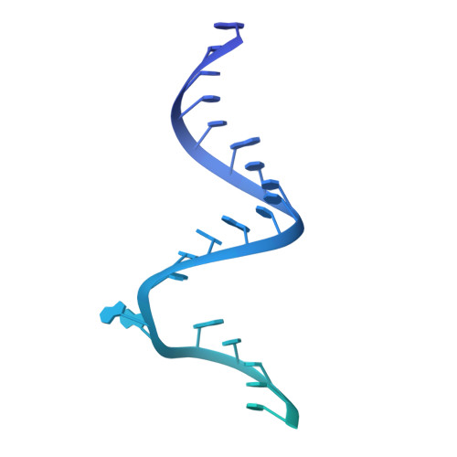
Structural basis for selective targeting of leishmanial ribosomes: aminoglycoside derivatives as promising therapeutics.
Shalev, M., Rozenberg, H., Smolkin, B., Nasereddin, A., Kopelyanskiy, D., Belakhov, V., Schrepfer, T., Schacht, J., Jaffe, C.L., Adir, N., Baasov, T.(2015) Nucleic Acids Res 43: 8601-8613
- PubMed: 26264664
- DOI: https://doi.org/10.1093/nar/gkv821
- Primary Citation of Related Structures:
4ZC7 - PubMed Abstract:
Leishmaniasis comprises an array of diseases caused by pathogenic species of Leishmania, resulting in a spectrum of mild to life-threatening pathologies. Currently available therapies for leishmaniasis include a limited selection of drugs. This coupled with the rather fast emergence of parasite resistance, presents a dire public health concern. Paromomycin (PAR), a broad-spectrum aminoglycoside antibiotic, has been shown in recent years to be highly efficient in treating visceral leishmaniasis (VL)-the life-threatening form of the disease. While much focus has been given to exploration of PAR activities in bacteria, its mechanism of action in Leishmania has received relatively little scrutiny and has yet to be fully deciphered. In the present study we present an X-ray structure of PAR bound to rRNA model mimicking its leishmanial binding target, the ribosomal A-site. We also evaluate PAR inhibitory actions on leishmanial growth and ribosome function, as well as effects on auditory sensory cells, by comparing several structurally related natural and synthetic aminoglycoside derivatives. The results provide insights into the structural elements important for aminoglycoside inhibitory activities and selectivity for leishmanial cytosolic ribosomes, highlighting a novel synthetic derivative, compound 3: , as a prospective therapeutic candidate for the treatment of VL.
- Schulich Faculty of Chemistry, Technion-Israel Institute of Technology, Haifa, Israel Department of Structural Biology, Faculty of Chemistry, Weizmann Institute of Science, Rehovot, Israel.
Organizational Affiliation:


