Crystal structure of O-methyltransferase CalO6 from the calicheamicin biosynthetic pathway: a case of challenging structure determination at low resolution.
Tsodikov, O.V., Hou, C., Walsh, C.T., Garneau-Tsodikova, S.(2015) BMC Struct Biol 15: 13-13
- PubMed: 26170207 Search on PubMedSearch on PubMed Central
- DOI: https://doi.org/10.1186/s12900-015-0040-6
- Primary Citation Related Structures:
4Z2Y - PubMed Abstract:
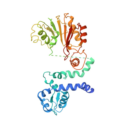
Calicheamicins (CAL) are enedyine natural products with potent antibiotic and cytotoxic activity, used in anticancer therapy. The O-methyltransferase CalO6 is proposed to catalyze methylation of the hydroxyl moiety at the C2 position of the orsellinic acid group of CAL. Crystals of CalO6 diffracted non-isotropically, with the usable data extending to 3.4 Å. While no single method of crystal structure determination yielded a structure of CalO6, we were able to determine its structure by using molecular replacement-guided single wavelength anomalous dispersion by using diffraction data from native crystals of CalO6 and a highly non-isomorphous mercury derivative. The structure of CalO6 reveals the methyltransferase fold and dimeric organization characteristic of small molecule O-methyltransferases involved in secondary metabolism in bacteria and plants. Uncommonly, CalO6 was crystallized in the absence of S-adenosylmethionine (SAM; the methyl donor) or S-adenosylhomocysteine (SAH; its product). Likely as a consequence of the dynamic nature of CalO6 in the absence of its cofactor, the central region of CalO6, which forms a helical lid-like structure near the active site in CalO6 and similar enzymes, is not observed in the electron density. We propose that this region controls the entry of SAM into and the exit of SAH from the active site of CalO6 and shapes the active site for substrate binding and catalysis.
- Department of Pharmaceutical Sciences, College of Pharmacy, University of Kentucky, 789 South Limestone Street, 40536-0596, Lexington, KY, USA. oleg.tsodikov@uky.edu.
Organizational Affiliation: