


Crystal structure of the tetrameric wt GluA2 ligand-binding domain bound to glutamate at 1.45 Angstroms resolution
Baranovic, J., Chebli, M., Salazar, H., Carbone, A.L., Ghisi, V., Faelber, K., Lau, A.Y., Daumke, O., Plested, A.J.R.To be published.
Experimental Data Snapshot
Starting Model: experimental
View more details
wwPDB Validation 3D Report Full Report

Entity ID: 1 | |||||
|---|---|---|---|---|---|
| Molecule | Chains | Sequence Length | Organism | Details | Image |
| Glutamate receptor 2,Glutamate receptor 2 | 263 | Rattus norvegicus | Mutation(s): 0 Gene Names: Gria2, Glur2 | 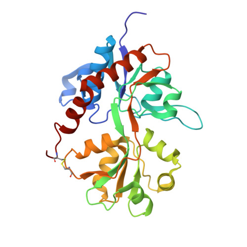 | |
UniProt | |||||
Entity Groups | |||||
| Sequence Clusters | 30% Identity50% Identity70% Identity90% Identity95% Identity100% Identity | ||||
| UniProt Group | P19491 | ||||
Sequence AnnotationsExpand | |||||
Reference Sequence | |||||
| Ligands 4 Unique | |||||
|---|---|---|---|---|---|
| ID | Chains | Name / Formula / InChI Key | 2D Diagram | 3D Interactions | |
| PG4 Download:Ideal Coordinates CCD File | G [auth A] | TETRAETHYLENE GLYCOL C8 H18 O5 UWHCKJMYHZGTIT-UHFFFAOYSA-N |  | ||
| GLU Download:Ideal Coordinates CCD File | C [auth A], I [auth B] | GLUTAMIC ACID C5 H9 N O4 WHUUTDBJXJRKMK-VKHMYHEASA-N |  | ||
| PEG Download:Ideal Coordinates CCD File | H [auth A] | DI(HYDROXYETHYL)ETHER C4 H10 O3 MTHSVFCYNBDYFN-UHFFFAOYSA-N |  | ||
| PO4 Download:Ideal Coordinates CCD File | D [auth A] E [auth A] F [auth A] J [auth B] K [auth B] | PHOSPHATE ION O4 P NBIIXXVUZAFLBC-UHFFFAOYSA-K |  | ||
| Length ( Å ) | Angle ( ˚ ) |
|---|---|
| a = 47.19 | α = 90 |
| b = 47.27 | β = 93.57 |
| c = 116.82 | γ = 90 |
| Software Name | Purpose |
|---|---|
| PHENIX | refinement |
| MOSFLM | data reduction |
| PHASER | phasing |
| Coot | model building |
| XDS | data reduction |
| XDS | data scaling |


