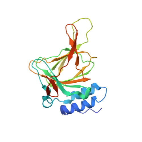
Influence of cysteine 164 on active site structure in rat cysteine dioxygenase.
Fellner, M., Siakkou, E., Faponle, A.S., Tchesnokov, E.P., de Visser, S.P., Wilbanks, S.M., Jameson, G.N.(2016) J Biol Inorg Chem 21: 501-510
- PubMed: 27193596 Search on PubMed
- DOI: https://doi.org/10.1007/s00775-016-1360-0
- Primary Citation Related Structures:
4YYO, 4Z82 - PubMed Abstract:
Cysteine dioxygenase is a non-heme mononuclear iron enzyme with unique structural features, namely an intramolecular thioether cross-link between cysteine 93 and tyrosine 157, and a disulfide bond between substrate L-cysteine and cysteine 164 in the entrance channel to the active site. We investigated how these posttranslational modifications affect catalysis through a kinetic, crystallographic and computational study. The enzyme kinetics of a C164S variant are identical to WT, indicating that disulfide formation at C164 does not significantly impair access to the active site at physiological pH. However, at high pH, the cysteine-tyrosine cross-link formation is enhanced in C164S. This supports the view that disulfide formation at position 164 can limit access to the active site. The C164S variant yielded crystal structures of unusual clarity in both resting state and with cysteine bound. Both show that the iron in the cysteine-bound complex is a mixture of penta- and hexa-coordinate with a water molecule taking up the final site (60 % occupancy), which is where dioxygen is believed to coordinate during turnover. The serine also displays stronger hydrogen bond interactions to a water bound to the amine of the substrate cysteine. However, the interactions between cysteine and iron appear unchanged. DFT calculations support this and show that WT and C164S have similar binding energies for the water molecule in the final site. This variant therefore provides evidence that WT also exists in an equilibrium between penta- and hexa-coordinate forms and the presence of the sixth ligand does not strongly affect dioxygen binding.
- Department of Chemistry, University of Otago, PO Box 56, Dunedin, 9054, New Zealand.
Organizational Affiliation: