4YUA | pdb_00004yua

Glycogen phosphorylase in complex with ellagic acid


Experimental Data Snapshot

  • Method: X-RAY DIFFRACTION
  • Resolution: 2.00 Å
  • R-Value Free: 
    0.202 (Depositor), 0.202 (DCC) 
  • R-Value Work: 
    0.173 (Depositor), 0.173 (DCC) 
  • R-Value Observed: 
    0.174 (Depositor) 

wwPDB Validation 3D Report Full Report

Validation slider image for 4YUA

Ligand Structure Quality Assessment 


This is version 2.1 of the entry. See complete history

Literature

Natural flavonoids as antidiabetic agents. The binding of gallic and ellagic acids to glycogen phosphorylase b.

Kyriakis, E.Stravodimos, G.A.Kantsadi, A.L.Chatzileontiadou, D.S.Skamnaki, V.T.Leonidas, D.D.

(2015) FEBS Lett 589: 1787-1794

  • DOI: https://doi.org/10.1016/j.febslet.2015.05.013
  • Primary Citation Related Structures: 
    4YUA, 4Z5X

  • PubMed Abstract: 

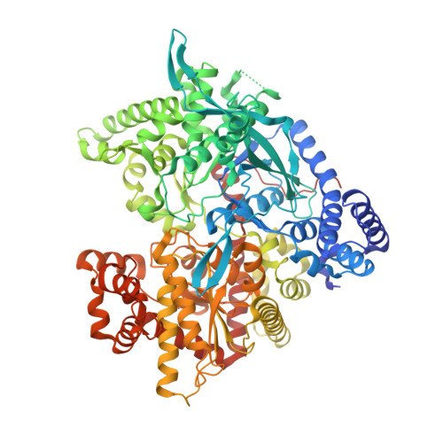
    We present a study on the binding of gallic acid and its dimer ellagic acid to glycogen phosphorylase (GP). Ellagic acid is a potent inhibitor with Kis of 13.4 and 7.5 μM, in contrast to gallic acid which displays Kis of 1.7 and 3.9 mM for GPb and GPa, respectively. Both compounds are competitive inhibitors with respect to the substrate, glucose-1-phoshate, and non-competitive to the allosteric activator, AMP. However, only ellagic acid functions with glucose in a strongly synergistic mode. The crystal structures of the GPb-gallic acid and GPb-ellagic acid complexes were determined at high resolution, revealing that both ligands bind to the inhibitor binding site of the enzyme and highlight the structural basis for the significant difference in their inhibitory potency.


  • Organizational Affiliation
    • Department of Biochemistry and Biotechnology, University of Thessaly, 26 Ploutonos Str., 41221 Larissa, Greece.

Macromolecule Content 

  • Total Structure Weight: 97.97 kDa 
  • Atom Count: 6,917 
  • Modeled Residue Count: 810 
  • Deposited Residue Count: 843 
  • Unique protein chains: 1

Macromolecules

Find similar proteins by:|  3D Structure
Entity ID: 1
MoleculeChains  Sequence LengthOrganismDetailsImage
Glycogen phosphorylase, muscle form843Oryctolagus cuniculusMutation(s): 0 
EC: 2.4.1.1
UniProt
Find proteins for P00489 (Oryctolagus cuniculus)
Explore P00489 
Go to UniProtKB:  P00489
Entity Groups
Sequence Clusters30% Identity50% Identity70% Identity90% Identity95% Identity100% Identity
UniProt GroupP00489
Sequence Annotations
Expand
Reference Sequence

Small Molecules

Ligands 2 Unique
IDChains Name / Formula / InChI Key2D Diagram3D Interactions
REF

Query on REF


Download:Ideal Coordinates CCD File
C [auth A]2,3,7,8-tetrahydroxychromeno[5,4,3-cde]chromene-5,10-dione
C14 H6 O8
AFSDNFLWKVMVRB-UHFFFAOYSA-N
PLP

Query on PLP


Download:Ideal Coordinates CCD File
B [auth A]PYRIDOXAL-5'-PHOSPHATE
C8 H10 N O6 P
NGVDGCNFYWLIFO-UHFFFAOYSA-N

Experimental Data & Validation

Experimental Data

  • Method: X-RAY DIFFRACTION
  • Resolution: 2.00 Å
  • R-Value Free:  0.202 (Depositor), 0.202 (DCC) 
  • R-Value Work:  0.173 (Depositor), 0.173 (DCC) 
  • R-Value Observed: 0.174 (Depositor) 
Space Group: P 43 21 2
Unit Cell:
Length ( Å )Angle ( ˚ )
a = 128.48α = 90
b = 128.48β = 90
c = 116.29γ = 90
Software Package:
Software NamePurpose
REFMACrefinement
MOSFLMdata reduction
SCALAdata scaling

Structure Validation

View Full Validation Report


Ligand Structure Quality Assessment 


Entry History 

Deposition Data

Revision History  (Full details and data files)

  • Version 1.0: 2015-05-13
    Type: Initial release
  • Version 1.1: 2015-06-03
    Changes: Database references
  • Version 1.2: 2015-07-15
    Changes: Database references
  • Version 2.0: 2018-01-17
    Changes: Atomic model, Data collection
  • Version 2.1: 2025-04-09
    Changes: Data collection, Database references, Structure summary