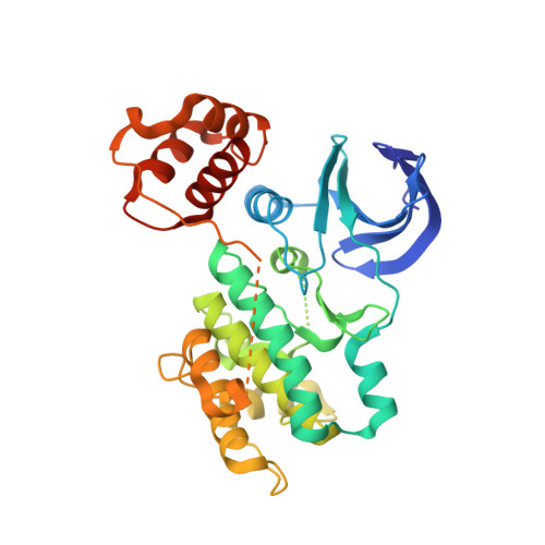
4YNZ

Structure of the N-terminal domain of SAD


Experimental Data Snapshot

  • Method: X-RAY DIFFRACTION
  • Resolution: 2.00 Å
  • R-Value Free: 0.229 
  • R-Value Work: 0.190 
  • R-Value Observed: 0.192 

Starting Model: experimental
View more details

wwPDB Validation   3D Report Full Report


This is version 1.2 of the entry. See complete history


Literature

Structural insight into the mechanism of synergistic autoinhibition of SAD kinases

Wu, J.X.Cheng, Y.S.Wang, J.Chen, L.Ding, M.Wu, J.W.

(2015) Nat Commun 6: 8953-8953

  • DOI: https://doi.org/10.1038/ncomms9953
  • Primary Citation of Related Structures:  
    4YNZ, 4YOM

  • PubMed Abstract: 

    The SAD/BRSK kinases participate in various important life processes, including neural development, cell cycle and energy metabolism. Like other members of the AMPK family, SAD contains an N-terminal kinase domain followed by the characteristic UBA and KA1 domains. Here we identify a unique autoinhibitory sequence (AIS) in SAD kinases, which exerts autoregulation in cooperation with UBA. Structural studies of mouse SAD-A revealed that UBA binds to the kinase domain in a distinct mode and, more importantly, AIS nestles specifically into the KD-UBA junction. The cooperative action of AIS and UBA results in an 'αC-out' inactive kinase, which is conserved across species and essential for presynaptic vesicle clustering in C. elegans. In addition, the AIS, along with the KA1 domain, is indispensable for phospholipid binding. Taken together, these data suggest a model for synergistic autoinhibition and membrane activation of SAD kinases.


  • Organizational Affiliation

    MOE Key Laboratory for Protein Science and Tsinghua-Peking Center for Life Sciences, School of Life Sciences, Tsinghua University, Beijing 100084, China.


Macromolecules
Find similar proteins by:  (by identity cutoff)  |  3D Structure
Entity ID: 1
MoleculeChains Sequence LengthOrganismDetailsImage
Serine/threonine-protein kinase BRSK2
A, B
342Mus musculusMutation(s): 0 
Gene Names: Brsk2
EC: 2.7.11.26
UniProt
Find proteins for Q69Z98 (Mus musculus)
Explore Q69Z98 
Go to UniProtKB:  Q69Z98
Entity Groups  
Sequence Clusters30% Identity50% Identity70% Identity90% Identity95% Identity100% Identity
UniProt GroupQ69Z98
Sequence Annotations
Expand
  • Reference Sequence
Experimental Data & Validation

Experimental Data

  • Method: X-RAY DIFFRACTION
  • Resolution: 2.00 Å
  • R-Value Free: 0.229 
  • R-Value Work: 0.190 
  • R-Value Observed: 0.192 
  • Space Group: P 1
Unit Cell:
Length ( Å )Angle ( ˚ )
a = 43.299α = 103.74
b = 60.745β = 106.48
c = 73.937γ = 107.42
Software Package:
Software NamePurpose
PHENIXrefinement
HKL-2000data collection
HKL-2000data scaling
PHASERphasing

Structure Validation

View Full Validation Report


Entry History & Funding Information

Deposition Data


Funding OrganizationLocationGrant Number
National Natural Science Foundation of ChinaChina31130062
National Natural Science Foundation of ChinaChina31321003
Ministry of Science and TechnologyChina2011CB910800
Ministry of Science and TechnologyChina2013CB530600

Revision History  (Full details and data files)

  • Version 1.0: 2015-12-16
    Type: Initial release
  • Version 1.1: 2017-10-18
    Changes: Author supporting evidence, Derived calculations
  • Version 1.2: 2023-11-08
    Changes: Data collection, Database references, Refinement description