4YLF | pdb_00004ylf

Insights into flavin-based electron bifurcation via the NADH-dependent reduced ferredoxin-NADP oxidoreductase structure


Experimental Data Snapshot

  • Method: X-RAY DIFFRACTION
  • Resolution: 2.30 Å
  • R-Value Free: 
    0.219 (Depositor), 0.229 (DCC) 
  • R-Value Work: 
    0.187 (Depositor) 
  • R-Value Observed: 
    0.191 (Depositor) 

wwPDB Validation 3D Report Full Report

Validation slider image for 4YLF

Ligand Structure Quality Assessment 


This is version 1.3 of the entry. See complete history

Literature

Insights into Flavin-based Electron Bifurcation via the NADH-dependent Reduced Ferredoxin:NADP Oxidoreductase Structure.

Demmer, J.K.Huang, H.Wang, S.Demmer, U.Thauer, R.K.Ermler, U.

(2015) J Biological Chem 290: 21985-21995

  • DOI: https://doi.org/10.1074/jbc.M115.656520
  • Primary Citation Related Structures: 
    4YLF, 4YRY

  • PubMed Abstract: 

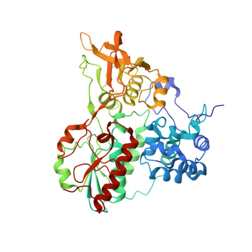
    NADH-dependent reduced ferredoxin:NADP oxidoreductase (NfnAB) is found in the cytoplasm of various anaerobic bacteria and archaea. The enzyme reversibly catalyzes the endergonic reduction of ferredoxin with NADPH driven by the exergonic transhydrogenation from NADPH onto NAD(+). Coupling is most probably accomplished via the mechanism of flavin-based electron bifurcation. To understand this process on a structural basis, we heterologously produced the NfnAB complex of Thermotoga maritima in Escherichia coli, provided kinetic evidence for its bifurcating behavior, and determined its x-ray structure in the absence and presence of NADH. The structure of NfnAB reveals an electron transfer route including the FAD (a-FAD), the [2Fe-2S] cluster of NfnA and the FAD (b-FAD), and the two [4Fe-4S] clusters of NfnB. Ferredoxin is presumably docked onto NfnB close to the [4Fe-4S] cluster distal to b-FAD. NAD(H) binds to a-FAD and NADP(H) consequently to b-FAD, which is positioned in the center of the NfnAB complex and the site of electron bifurcation. Arg(187) is hydrogen-bonded to N5 and O4 of the bifurcating b-FAD and might play a key role in adjusting a low redox potential of the FADH(•)/FAD pair required for ferredoxin reduction. A mechanism of FAD-coupled electron bifurcation by NfnAB is proposed.


  • Organizational Affiliation
    • From the Max-Planck-Institut für Biophysik, D-60438 Frankfurt am Main, Germany and the Max-Planck-Institut für Terrestrische Mikrobiologie, D-35043 Marburg, Germany.

Macromolecule Content 

  • Total Structure Weight: 169.57 kDa 
  • Atom Count: 11,843 
  • Modeled Residue Count: 1,475 
  • Deposited Residue Count: 1,488 
  • Unique protein chains: 2

Macromolecules

Find similar proteins by:|  3D Structure
Entity ID: 1
MoleculeChains  Sequence LengthOrganismDetailsImage
Dihydroorotate dehydrogenase B (NAD(+)), electron transfer subunit homolog
A, C
276Thermotoga maritima MSB8Mutation(s): 0 
Gene Names: TM_1639
UniProt
Find proteins for Q9X1X4 (Thermotoga maritima (strain ATCC 43589 / DSM 3109 / JCM 10099 / NBRC 100826 / MSB8))
Explore Q9X1X4 
Go to UniProtKB:  Q9X1X4
Entity Groups
Sequence Clusters30% Identity50% Identity70% Identity90% Identity95% Identity100% Identity
UniProt GroupQ9X1X4
Sequence Annotations
Expand
Reference Sequence
Find similar proteins by:|  3D Structure
Entity ID: 2
MoleculeChains  Sequence LengthOrganismDetailsImage
Dihydropyrimidine dehydrogenase subunit A
B, D
468Thermotoga maritima MSB8Mutation(s): 0 
Gene Names: THEMA_06045
UniProt
Find proteins for Q9X1X5 (Thermotoga maritima (strain ATCC 43589 / DSM 3109 / JCM 10099 / NBRC 100826 / MSB8))
Explore Q9X1X5 
Go to UniProtKB:  Q9X1X5
Entity Groups
Sequence Clusters30% Identity50% Identity70% Identity90% Identity95% Identity100% Identity
UniProt GroupQ9X1X5
Sequence Annotations
Expand
Reference Sequence

Small Molecules

Ligands 4 Unique
IDChains Name / Formula / InChI Key2D Diagram3D Interactions
FAD

Query on FAD


Download:Ideal Coordinates CCD File
F [auth A],
I [auth B],
K [auth C],
O [auth D]
FLAVIN-ADENINE DINUCLEOTIDE
C27 H33 N9 O15 P2
VWWQXMAJTJZDQX-UYBVJOGSSA-N
SF4

Query on SF4


Download:Ideal Coordinates CCD File
G [auth B],
H [auth B],
M [auth D],
N [auth D]
IRON/SULFUR CLUSTER
Fe4 S4
LJBDFODJNLIPKO-UHFFFAOYSA-N
FES

Query on FES


Download:Ideal Coordinates CCD File
E [auth A],
L [auth C]
FE2/S2 (INORGANIC) CLUSTER
Fe2 S2
NIXDOXVAJZFRNF-UHFFFAOYSA-N
PO4

Query on PO4


Download:Ideal Coordinates CCD File
J [auth B],
P [auth D]
PHOSPHATE ION
O4 P
NBIIXXVUZAFLBC-UHFFFAOYSA-K

Experimental Data & Validation

Experimental Data

  • Method: X-RAY DIFFRACTION
  • Resolution: 2.30 Å
  • R-Value Free:  0.219 (Depositor), 0.229 (DCC) 
  • R-Value Work:  0.187 (Depositor) 
  • R-Value Observed: 0.191 (Depositor) 
Space Group: P 43
Unit Cell:
Length ( Å )Angle ( ˚ )
a = 81.42α = 90
b = 81.42β = 90
c = 311.48γ = 90
Software Package:
Software NamePurpose
PHENIXrefinement
XSCALEdata scaling
SHARPphasing
XDSdata reduction

Structure Validation

View Full Validation Report


Ligand Structure Quality Assessment 


Entry History 

Deposition Data

Revision History  (Full details and data files)

  • Version 1.0: 2015-07-08
    Type: Initial release
  • Version 1.1: 2015-07-15
    Changes: Database references
  • Version 1.2: 2015-09-16
    Changes: Database references
  • Version 1.3: 2024-05-08
    Changes: Data collection, Database references