4YIF

Crystal structure of Rv0880


Experimental Data Snapshot

  • Method: X-RAY DIFFRACTION
  • Resolution: 2.00 Å
  • R-Value Free: 0.249 
  • R-Value Work: 0.212 
  • R-Value Observed: 0.214 

wwPDB Validation   3D Report Full Report


This is version 1.3 of the entry. See complete history


Literature

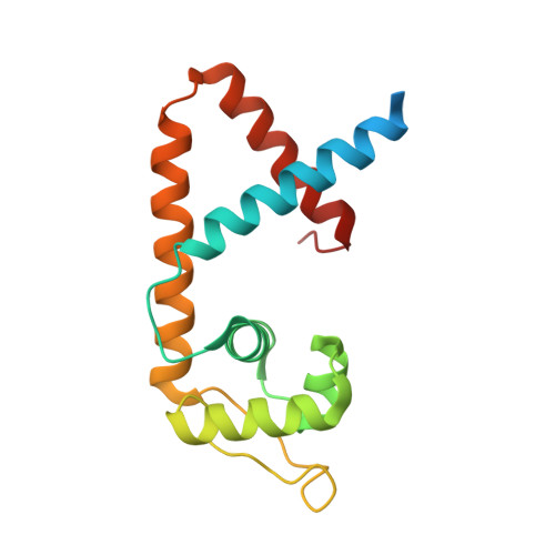
Structure of the MarR family protein Rv0880 from Mycobacterium tuberculosis.

Gao, Y.R.Feng, N.Chen, T.Li, D.F.Bi, L.J.

(2015) Acta Crystallogr F Struct Biol Commun 71: 741-745

  • DOI: https://doi.org/10.1107/S2053230X15007281
  • Primary Citation of Related Structures:  
    4YIF

  • PubMed Abstract: 

    Rv0880 from the pathogen Mycobacterium tuberculosis is classified as a MarR family protein in the Pfam database. It consists of 143 amino acids and has an isoelectric point of 10.9. Crystals of Rv0880 belonged to space group P1, with unit-cell parameters a = 54.97, b = 69.60, c = 70.32 Å, α = 103.71, β = 111.06, γ = 105.83°. The structure of the MarR family transcription regulator Rv0880 was solved at a resolution of 2.0 Å with an R(cryst) and R(free) of 21.2 and 24.9%, respectively. The dimeric structure resembles that of other MarR proteins, with each subunit comprising a winged helix-turn-helix domain connected to an α-helical dimerization domain.


  • Organizational Affiliation

    Key Laboratory of RNA Biology, National Laboratory of Biomacromolecules, Institute of Biophysics, Chinese Academy of Sciences, 15 Datun Road, Beijing 100101, People's Republic of China.


Macromolecules
Find similar proteins by:  (by identity cutoff)  |  3D Structure
Entity ID: 1
MoleculeChains Sequence LengthOrganismDetailsImage
MarR family protein Rv0880
A, B, C, D, E
A, B, C, D, E, F
164Mycobacterium tuberculosis H37RvMutation(s): 0 
Gene Names: Rv0880MTCY31.08
UniProt
Find proteins for P9WMF1 (Mycobacterium tuberculosis (strain ATCC 25618 / H37Rv))
Explore P9WMF1 
Go to UniProtKB:  P9WMF1
Entity Groups  
Sequence Clusters30% Identity50% Identity70% Identity90% Identity95% Identity100% Identity
UniProt GroupP9WMF1
Sequence Annotations
Expand
  • Reference Sequence
Experimental Data & Validation

Experimental Data

  • Method: X-RAY DIFFRACTION
  • Resolution: 2.00 Å
  • R-Value Free: 0.249 
  • R-Value Work: 0.212 
  • R-Value Observed: 0.214 
  • Space Group: P 1
Unit Cell:
Length ( Å )Angle ( ˚ )
a = 54.97α = 103.71
b = 69.6β = 111.06
c = 70.32γ = 105.83
Software Package:
Software NamePurpose
PHENIXrefinement
MOSFLMdata reduction
SCALAdata scaling
MR-Rosettaphasing

Structure Validation

View Full Validation Report


Entry History & Funding Information

Deposition Data


Funding OrganizationLocationGrant Number
Ministry of Science and Technology (China)China2011CB910302

Revision History  (Full details and data files)

  • Version 1.0: 2015-06-10
    Type: Initial release
  • Version 1.1: 2017-10-18
    Changes: Author supporting evidence, Derived calculations, Source and taxonomy, Structure summary
  • Version 1.2: 2019-12-25
    Changes: Database references
  • Version 1.3: 2023-11-08
    Changes: Data collection, Database references, Refinement description