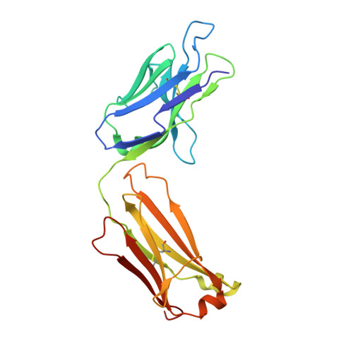
4YHO

Reversal Agent for Dabigatran


Experimental Data Snapshot

  • Method: X-RAY DIFFRACTION
  • Resolution: 1.82 Å
  • R-Value Free: 0.210 
  • R-Value Work: 0.180 
  • R-Value Observed: 0.182 

Starting Model: experimental
View more details

wwPDB Validation   3D Report Full Report


Ligand Structure Quality Assessment 


This is version 1.1 of the entry. See complete history


Literature

Structure-guided residence time optimization of a dabigatran reversal agent.

Schiele, F.van Ryn, J.Litzenburger, T.Ritter, M.Seeliger, D.Nar, H.

(2015) MAbs 7: 871-880

  • DOI: https://doi.org/10.1080/19420862.2015.1057364
  • Primary Citation of Related Structures:  
    4YGV, 4YHI, 4YHK, 4YHL, 4YHM, 4YHN, 4YHO

  • PubMed Abstract: 

    Novel oral anticoagulants are effective and safe alternatives to vitamin-K antagonists for anticoagulation therapy. However, anticoagulation therapy in general is associated with an elevated risk of bleeding. Idarucizumab is a reversal agent for the direct thrombin inhibitor, dabigatran etexilate (Pradaxa®) and is currently in Phase 3 studies. Here, we report data on the antibody fragment aDabi-Fab2, a putative backup molecule for idarucizumab. Although aDabi-Fab2 completely reversed effects of dabigatran in a rat model in vivo, we observed significantly reduced duration of action compared to idarucizumab. Rational protein engineering, based on the X-ray structure of aDabi-Fab2, led to the identification of mutant Y103W. The mutant had optimized shape complementarity to dabigatran while maintaining an energetically favored hydrogen bond. It displayed increased affinity for dabigatran, mainly driven by a slower off-rate. Interestingly, the increased residence time translated into longer duration of action in vivo. It was thus possible to further enhance the efficacy of aDabi-Fab2 based on rational design, giving it the potential to serve as a back-up candidate for idarucizumab.


  • Organizational Affiliation

    a New Biological Entities Discovery; Boehringer Ingelheim GmbH & Co. KG ; Biberach , Germany.


Macromolecules
Find similar proteins by:  (by identity cutoff)  |  3D Structure
Entity ID: 1
MoleculeChains Sequence LengthOrganismDetailsImage
aDabi-Fab3 heavy chainA [auth H]222Homo sapiensMutation(s): 0 
Entity Groups  
Sequence Clusters30% Identity50% Identity70% Identity90% Identity95% Identity100% Identity
Sequence Annotations
Expand
  • Reference Sequence
Find similar proteins by:  (by identity cutoff)  |  3D Structure
Entity ID: 2
MoleculeChains Sequence LengthOrganismDetailsImage
aDabi-Fab3 light chainB [auth L]219Homo sapiensMutation(s): 0 
Entity Groups  
Sequence Clusters30% Identity50% Identity70% Identity90% Identity95% Identity100% Identity
Sequence Annotations
Expand
  • Reference Sequence
Small Molecules
Ligands 1 Unique
IDChains Name / Formula / InChI Key2D Diagram3D Interactions
4CC
Query on 4CC

Download Ideal Coordinates CCD File 
C [auth H]N-[(2-{[(4-carbamimidoylphenyl)amino]methyl}-1-methyl-1H-benzimidazol-5-yl)carbonyl]-N-pyridin-2-yl-beta-alanine
C25 H25 N7 O3
YBSJFWOBGCMAKL-UHFFFAOYSA-N
Experimental Data & Validation

Experimental Data

  • Method: X-RAY DIFFRACTION
  • Resolution: 1.82 Å
  • R-Value Free: 0.210 
  • R-Value Work: 0.180 
  • R-Value Observed: 0.182 
  • Space Group: P 21 21 21
Unit Cell:
Length ( Å )Angle ( ˚ )
a = 60.278α = 90
b = 78.089β = 90
c = 93.372γ = 90
Software Package:
Software NamePurpose
BUSTERrefinement
XDSdata reduction
XDSdata scaling
BUSTER-TNTphasing

Structure Validation

View Full Validation Report


Ligand Structure Quality Assessment 


Entry History 

Deposition Data

Revision History  (Full details and data files)

  • Version 1.0: 2016-01-13
    Type: Initial release
  • Version 1.1: 2024-01-10
    Changes: Data collection, Database references, Refinement description