


Structure of homoisocitrate dehydrogenase from Thermus thermophilus in complex with homoisocitrate, magnesium(II) and NADH
Takahashi, K., Tomita, T., Kuzuyama, T., Nishiyama, M.To be published.
Experimental Data Snapshot
Starting Model: experimental
View more details
Entity ID: 1 | |||||
|---|---|---|---|---|---|
| Molecule | Chains | Sequence Length | Organism | Details | Image |
| Homoisocitrate dehydrogenase | 334 | Thermus thermophilus HB27 | Mutation(s): 0 Gene Names: hicd, hdh, hicdh, TT_C1012 EC: 1.1.1.87 (PDB Primary Data), 1.1.1.286 (UniProt) | 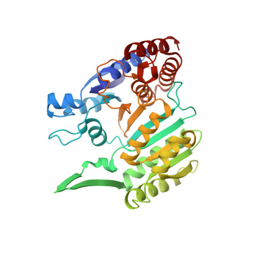 | |
UniProt | |||||
Entity Groups | |||||
| Sequence Clusters | 30% Identity50% Identity70% Identity90% Identity95% Identity100% Identity | ||||
| UniProt Group | Q72IW9 | ||||
Sequence AnnotationsExpand | |||||
Reference Sequence | |||||
| Ligands 5 Unique | |||||
|---|---|---|---|---|---|
| ID | Chains | Name / Formula / InChI Key | 2D Diagram | 3D Interactions | |
| NAI Download:Ideal Coordinates CCD File | G [auth A], GB [auth D], LA [auth C], U [auth B] | 1,4-DIHYDRONICOTINAMIDE ADENINE DINUCLEOTIDE C21 H29 N7 O14 P2 BOPGDPNILDQYTO-NNYOXOHSSA-N |  | ||
| 48Y Download:Ideal Coordinates CCD File | F [auth A], FB [auth D], KA [auth C], T [auth B] | (1R,2S)-1-hydroxybutane-1,2,4-tricarboxylic acid C7 H10 O7 OEJZZCGRGVFWHK-WVZVXSGGSA-N |  | ||
| SO4 Download:Ideal Coordinates CCD File | AB [auth C] BA [auth B] BB [auth C] CA [auth B] CB [auth C] | SULFATE ION O4 S QAOWNCQODCNURD-UHFFFAOYSA-L |  | ||
| GOL Download:Ideal Coordinates CCD File | AA [auth B] HB [auth D] I [auth A] IB [auth D] J [auth A] | GLYCEROL C3 H8 O3 PEDCQBHIVMGVHV-UHFFFAOYSA-N |  | ||
| MG Download:Ideal Coordinates CCD File | E [auth A], H [auth A], JA [auth C], MA [auth C] | MAGNESIUM ION Mg JLVVSXFLKOJNIY-UHFFFAOYSA-N |  | ||
| Length ( Å ) | Angle ( ˚ ) |
|---|---|
| a = 159.663 | α = 90 |
| b = 159.663 | β = 90 |
| c = 148.517 | γ = 90 |
| Software Name | Purpose |
|---|---|
| REFMAC | refinement |
| Coot | refinement |
| HKL-2000 | data scaling |
| PHASER | phasing |


