Structural Basis for Iron-Mediated Sulfur Transfer in Archael and Yeast Thiazole Synthases.
Zhang, X., Eser, B.E., Chanani, P.K., Begley, T.P., Ealick, S.E.(2016) Biochemistry 55: 1826-1838
- PubMed: 26919468 Search on PubMedSearch on PubMed Central
- DOI: https://doi.org/10.1021/acs.biochem.6b00030
- Primary Citation Related Structures:
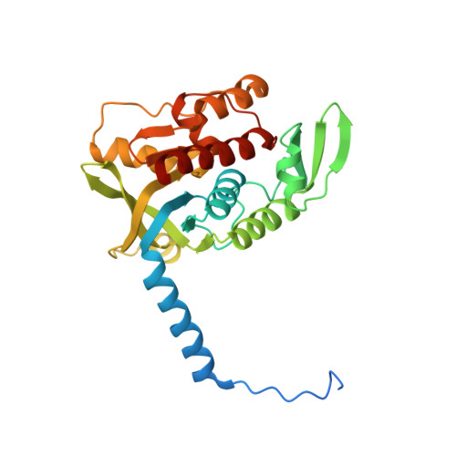
4Y4L, 4Y4M, 4Y4N - PubMed Abstract:
Thiamin diphosphate is an essential cofactor in all forms of life and plays a key role in amino acid and carbohydrate metabolism. Its biosynthesis involves separate syntheses of the pyrimidine and thiazole moieties, which are then coupled to form thiamin monophosphate. A final phosphorylation produces the active form of the cofactor. In most bacteria, six gene products are required for biosynthesis of the thiamin thiazole. In yeast and fungi only one gene product, Thi4, is required for thiazole biosynthesis. Methanococcus jannaschii expresses a putative Thi4 ortholog that was previously reported to be a ribulose 1,5-bisphosphate synthase [Finn, M. W. and Tabita, F. R. (2004) J. Bacteriol., 186, 6360-6366]. Our structural studies show that the Thi4 orthologs from M. jannaschii and Methanococcus igneus are structurally similar to Thi4 from Saccharomyces cerevisiae. In addition, all active site residues are conserved except for a key cysteine residue, which in S. cerevisiae is the source of the thiazole sulfur atom. Our recent biochemical studies showed that the archael Thi4 orthologs use nicotinamide adenine dinucleotide, glycine, and free sulfide to form the thiamin thiazole in an iron-dependent reaction [Eser, B., Zhang, X., Chanani, P. K., Begley, T. P., and Ealick, S. E. (2016) J. Am. Chem. Soc. , DOI: 10.1021/jacs.6b00445]. Here we report X-ray crystal structures of Thi4 from M. jannaschii complexed with ADP-ribulose, the C205S variant of Thi4 from S. cerevisiae with a bound glycine imine intermediate, and Thi4 from M. igneus with bound glycine imine intermediate and iron. These studies reveal the structural basis for the iron-dependent mechanism of sulfur transfer in archael and yeast thiazole synthases.
- Department of Chemistry and Chemical Biology, Cornell University , Ithaca, New York 14853, United States.
Organizational Affiliation: