4Y1S | pdb_00004y1s

Structural basis for Ca2+-mediated interaction of the perforin C2 domain with lipid membranes


Experimental Data Snapshot

  • Method: X-RAY DIFFRACTION
  • Resolution: 1.61 Å
  • R-Value Free: 
    0.185 (Depositor), 0.195 (DCC) 
  • R-Value Work: 
    0.166 (Depositor) 
  • R-Value Observed: 
    0.167 (Depositor) 

Starting Model: experimental
View more details

wwPDB Validation 3D Report Full Report

Validation slider image for 4Y1S

This is version 1.5 of the entry. See complete history

Literature

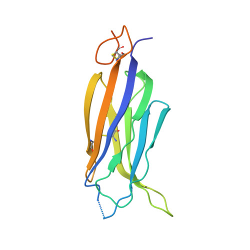
Structural Basis for Ca2+-mediated Interaction of the Perforin C2 Domain with Lipid Membranes.

Yagi, H.Conroy, P.J.Leung, E.W.Law, R.H.Trapani, J.A.Voskoboinik, I.Whisstock, J.C.Norton, R.S.

(2015) J Biological Chem 290: 25213-25226

  • DOI: https://doi.org/10.1074/jbc.M115.668384
  • Primary Citation Related Structures: 
    4Y1S, 4Y1T

  • PubMed Abstract: 

    Natural killer cells and cytotoxic T-lymphocytes deploy perforin and granzymes to kill infected host cells. Perforin, secreted by immune cells, binds target membranes to form pores that deliver pro-apoptotic granzymes into the target cell. A crucial first step in this process is interaction of its C2 domain with target cell membranes, which is a calcium-dependent event. Some aspects of this process are understood, but many molecular details remain unclear. To address this, we investigated the mechanism of Ca(2+) and lipid binding to the C2 domain by NMR spectroscopy and x-ray crystallography. Calcium titrations, together with dodecylphosphocholine micelle experiments, confirmed that multiple Ca(2+) ions bind within the calcium-binding regions, activating perforin with respect to membrane binding. We have also determined the affinities of several of these binding sites and have shown that this interaction causes a significant structural rearrangement in CBR1. Thus, it is proposed that Ca(2+) binding at the weakest affinity site triggers changes in the C2 domain that facilitate its interaction with lipid membranes.


  • Organizational Affiliation
    • From the Monash Institute of Pharmaceutical Sciences, Monash University, Parkville, Victoria 3052.

Macromolecule Content 

  • Total Structure Weight: 16.52 kDa 
  • Atom Count: 1,120 
  • Modeled Residue Count: 122 
  • Deposited Residue Count: 149 
  • Unique protein chains: 1

Macromolecules

Find similar proteins by:|  3D Structure
Entity ID: 1
MoleculeChains  Sequence LengthOrganismDetailsImage
Perforin-1149Mus musculusMutation(s): 4 
Gene Names: Prf1Pfp
UniProt & NIH Common Fund Data Resources
Find proteins for P10820 (Mus musculus)
Explore P10820 
Go to UniProtKB:  P10820
IMPC:  MGI:97551
Entity Groups
Sequence Clusters30% Identity50% Identity70% Identity90% Identity95% Identity100% Identity
UniProt GroupP10820
Sequence Annotations
Expand
Reference Sequence

Small Molecules

Ligands 1 Unique
IDChains Name / Formula / InChI Key2D Diagram3D Interactions
CA

Query on CA


Download:Ideal Coordinates CCD File
B [auth A]CALCIUM ION
Ca
BHPQYMZQTOCNFJ-UHFFFAOYSA-N

Experimental Data & Validation

Experimental Data

  • Method: X-RAY DIFFRACTION
  • Resolution: 1.61 Å
  • R-Value Free:  0.185 (Depositor), 0.195 (DCC) 
  • R-Value Work:  0.166 (Depositor) 
  • R-Value Observed: 0.167 (Depositor) 
Space Group: P 21 21 21
Unit Cell:
Length ( Å )Angle ( ˚ )
a = 37.7α = 90
b = 43.211β = 90
c = 68.077γ = 90
Software Package:
Software NamePurpose
PHENIXrefinement
XDSdata reduction
Blu-Icedata collection
SCALAdata scaling
PHASERphasing

Structure Validation

View Full Validation Report


Entry History 

Deposition Data

Revision History  (Full details and data files)

  • Version 1.0: 2015-09-02
    Type: Initial release
  • Version 1.1: 2015-09-09
    Changes: Database references
  • Version 1.2: 2015-10-28
    Changes: Database references
  • Version 1.3: 2017-12-13
    Changes: Data collection, Database references, Derived calculations, Source and taxonomy
  • Version 1.4: 2023-09-27
    Changes: Data collection, Database references, Derived calculations, Refinement description
  • Version 1.5: 2024-11-06
    Changes: Structure summary