4XXW

Crystal structure of mouse Cadherin-23 EC1-2 and Protocadherin-15 EC1-2 splice variant


Experimental Data Snapshot

  • Method: X-RAY DIFFRACTION
  • Resolution: 2.26 Å
  • R-Value Free: 0.233 
  • R-Value Work: 0.187 
  • R-Value Observed: 0.189 

wwPDB Validation   3D Report Full Report


This is version 1.4 of the entry. See complete history


Literature

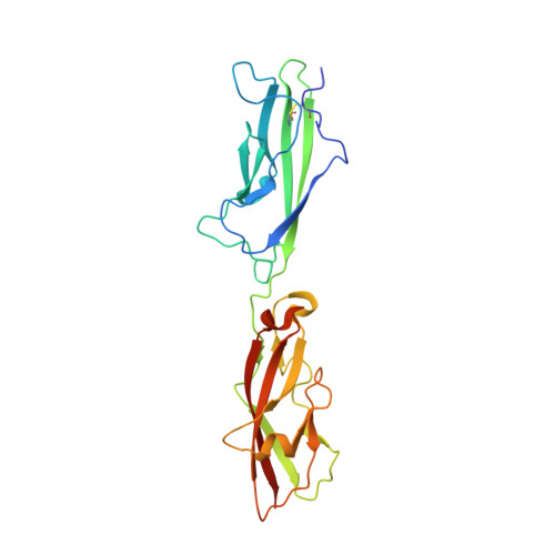
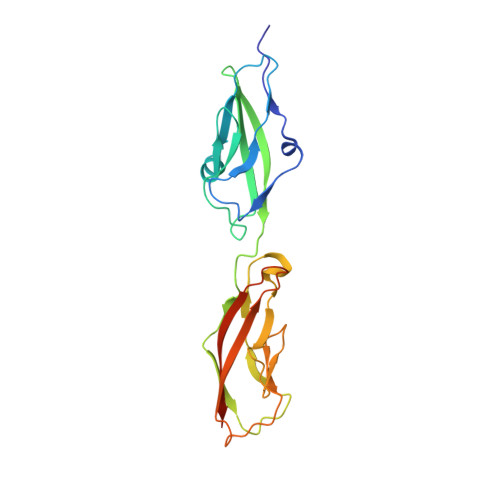
Tuning Inner-Ear Tip-Link Affinity Through Alternatively Spliced Variants of Protocadherin-15.

Narui, Y.Sotomayor, M.

(2018) Biochemistry 57: 1702-1710

  • DOI: https://doi.org/10.1021/acs.biochem.7b01075
  • Primary Citation of Related Structures:  
    4XXW

  • PubMed Abstract: 

    Human hearing relies upon the tip-to-tip interaction of two nonclassical cadherins, protocadherin-15 (PCDH15) and cadherin-23 (CDH23). Together, these proteins form a filament called the tip link that connects neighboring stereocilia of mechanosensitive hair cells. As sound waves enter the cochlea, the stereocilia deflect and tension is applied to the tip link, opening nearby transduction channels. Disruption of the tip link by loud sound or calcium chelators eliminates transduction currents and illustrates that tip-link integrity is critical for mechanosensing. Tip-link remodeling after disruption is a dynamic process, which can lead to the formation of atypical complexes that incorporate alternatively spliced variants of PCDH15. These variants are categorized into six groups (N1-N6) based upon differences in the first two extracellular cadherin (EC) repeats. Here, we characterized the two N-terminal EC repeats of all PCDH15 variants (pcdh15(N1) to pcdh15(N6)) and combined these variants to test complex formation. We solved the crystal structure of a new complex composed of CDH23 EC1-2 (cdh23) and pcdh15(N2) at 2.3 Å resolution and compared it to the canonical cdh23-pcdh15(N1) complex. While there were subtle structural differences, the binding affinity between cdh23 and pcdh15(N2) is ∼6 times weaker than cdh23 and pcdh15(N1) as determined by surface plasmon resonance analysis. Steered molecular dynamics simulations predict that the unbinding force of the cdh23-pcdh15(N2) complex can be lower than the canonical tip link. Our results demonstrate that alternative heterophilic tip-link structures form stable protein-protein interactions in vitro and suggest that homophilic PCDH15-PCDH15 tip links form through the interaction of additional EC repeats.


  • Organizational Affiliation

    Department of Chemistry and Biochemistry , The Ohio State University , Columbus , Ohio 43210 , United States.


Macromolecules
Find similar proteins by:  (by identity cutoff)  |  3D Structure
Entity ID: 1
MoleculeChains Sequence LengthOrganismDetailsImage
Protocadherin-15A [auth B],
B [auth A]
237Mus musculusMutation(s): 0 
Gene Names: Pcdh15
UniProt & NIH Common Fund Data Resources
Find proteins for Q99PJ1 (Mus musculus)
Explore Q99PJ1 
Go to UniProtKB:  Q99PJ1
IMPC:  MGI:1891428
Entity Groups  
Sequence Clusters30% Identity50% Identity70% Identity90% Identity95% Identity100% Identity
UniProt GroupQ99PJ1
Sequence Annotations
Expand
  • Reference Sequence
Find similar proteins by:  (by identity cutoff)  |  3D Structure
Entity ID: 2
MoleculeChains Sequence LengthOrganismDetailsImage
Cadherin-23
C, D
214Mus musculusMutation(s): 0 
Gene Names: Cdh23
UniProt & NIH Common Fund Data Resources
Find proteins for Q99PF4 (Mus musculus)
Explore Q99PF4 
Go to UniProtKB:  Q99PF4
IMPC:  MGI:1890219
Entity Groups  
Sequence Clusters30% Identity50% Identity70% Identity90% Identity95% Identity100% Identity
UniProt GroupQ99PF4
Sequence Annotations
Expand
  • Reference Sequence
Small Molecules
Ligands 2 Unique
IDChains Name / Formula / InChI Key2D Diagram3D Interactions
CA
Query on CA

Download Ideal Coordinates CCD File 
E [auth B]
F [auth B]
G [auth B]
I [auth A]
J [auth A]
E [auth B],
F [auth B],
G [auth B],
I [auth A],
J [auth A],
K [auth A],
M [auth C],
N [auth C],
O [auth C],
P [auth C],
Q [auth D],
R [auth D],
S [auth D],
T [auth D]
CALCIUM ION
Ca
BHPQYMZQTOCNFJ-UHFFFAOYSA-N
CL
Query on CL

Download Ideal Coordinates CCD File 
H [auth B],
L [auth A]
CHLORIDE ION
Cl
VEXZGXHMUGYJMC-UHFFFAOYSA-M
Experimental Data & Validation

Experimental Data

Unit Cell:
Length ( Å )Angle ( ˚ )
a = 157.746α = 90
b = 57.789β = 99.17
c = 155.264γ = 90
Software Package:
Software NamePurpose
REFMACrefinement
HKL-2000data reduction
HKL-2000data scaling
PHASERphasing

Structure Validation

View Full Validation Report


Entry History & Funding Information

Deposition Data


Funding OrganizationLocationGrant Number
National Institutes of Health/National Institute on Deafness and Other Communication Disorders (NIH/NIDCD)United StatesR00 DC012534

Revision History  (Full details and data files)

  • Version 1.0: 2016-05-04
    Type: Initial release
  • Version 1.1: 2017-09-06
    Changes: Author supporting evidence, Derived calculations
  • Version 1.2: 2018-08-01
    Changes: Data collection, Database references
  • Version 1.3: 2019-12-18
    Changes: Author supporting evidence
  • Version 1.4: 2023-09-27
    Changes: Data collection, Database references, Derived calculations, Refinement description