4XO2

crystal structure of GnsA from E.coli


Experimental Data Snapshot

  • Method: X-RAY DIFFRACTION
  • Resolution: 1.95 Å
  • R-Value Free: 0.250 
  • R-Value Work: 0.213 
  • R-Value Observed: 0.217 

wwPDB Validation   3D Report Full Report


This is version 1.1 of the entry. See complete history


Literature

Crystal structure of GnsA from Escherichia coli

Wei, Y.Zhan, L.Gao, Z.Prive, G.G.Dong, Y.

(2015) Biochem Biophys Res Commun 462: 1-7

  • DOI: https://doi.org/10.1016/j.bbrc.2015.03.133
  • Primary Citation of Related Structures:  
    4XO1, 4XO2

  • PubMed Abstract: 

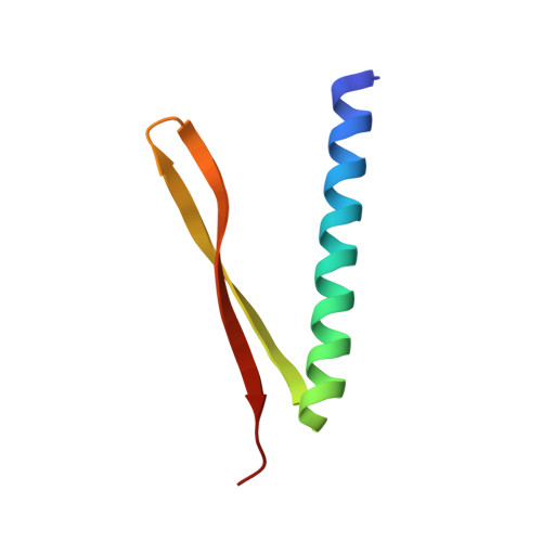
    Escherichia Coli GnsA is a regulator of phosphatidylethanolamine synthesis and functions as a suppressor of both a secG null mutation and fabA6 mutations. GnsA may also be a toxin with the cognate antitoxin YmcE. Here we report the crystal structure of GnsA to 1.8 Å. GnsA forms a V shaped hairpin structure that is tightly associated into a homodimer. Our comprehensive structural study suggests that GnsA is structurally similar to an outer membrane protein, suggesting a function of protein binding.


  • Organizational Affiliation

    School of Life Science, University of Science and Technology of China, Hefei 230027, China; Department of Medical Biophysics, University of Toronto, Toronto, Ontario, Canada. Electronic address: yongwei@uhnresearch.ca.


Macromolecules
Find similar proteins by:  (by identity cutoff)  |  3D Structure
Entity ID: 1
MoleculeChains Sequence LengthOrganismDetailsImage
Protein GnsA
A, B
60Escherichia coli K-12Mutation(s): 0 
Gene Names: gnsAyccLb4517JW0976
UniProt
Find proteins for P0AC92 (Escherichia coli (strain K12))
Explore P0AC92 
Go to UniProtKB:  P0AC92
Entity Groups  
Sequence Clusters30% Identity50% Identity70% Identity90% Identity95% Identity100% Identity
UniProt GroupP0AC92
Sequence Annotations
Expand
  • Reference Sequence
Experimental Data & Validation

Experimental Data

  • Method: X-RAY DIFFRACTION
  • Resolution: 1.95 Å
  • R-Value Free: 0.250 
  • R-Value Work: 0.213 
  • R-Value Observed: 0.217 
  • Space Group: C 1 2 1
Unit Cell:
Length ( Å )Angle ( ˚ )
a = 42.532α = 90
b = 38.047β = 90.1
c = 72.626γ = 90
Software Package:
Software NamePurpose
PHENIXrefinement
HKL-2000data reduction
HKL-2000data scaling
PHASERphasing

Structure Validation

View Full Validation Report


Entry History 

Deposition Data

Revision History  (Full details and data files)

  • Version 1.0: 2015-06-17
    Type: Initial release
  • Version 1.1: 2024-03-20
    Changes: Data collection, Database references, Derived calculations, Source and taxonomy, Structure summary