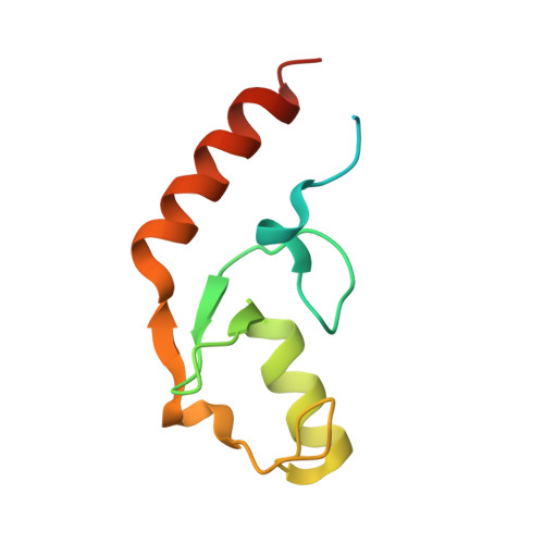
4XI1

Crystal structure of U-box 2 of LubX / LegU2 / Lpp2887 from Legionella pneumophila str. Paris, wild-type


Experimental Data Snapshot

  • Method: X-RAY DIFFRACTION
  • Resolution: 2.98 Å
  • R-Value Free: 0.209 
  • R-Value Work: 0.162 
  • R-Value Observed: 0.165 

Starting Model: experimental
View more details

wwPDB Validation   3D Report Full Report


This is version 1.3 of the entry. See complete history


Literature

Molecular Characterization of LubX: Functional Divergence of the U-Box Fold by Legionella pneumophila.

Quaile, A.T.Urbanus, M.L.Stogios, P.J.Nocek, B.Skarina, T.Ensminger, A.W.Savchenko, A.

(2015) Structure 23: 1459-1469

  • DOI: https://doi.org/10.1016/j.str.2015.05.020
  • Primary Citation of Related Structures:  
    4WZ0, 4WZ2, 4WZ3, 4XI1

  • PubMed Abstract: 

    LubX is part of the large arsenal of effectors in Legionella pneumophila that are translocated into the host cytosol during infection. Despite such unique features as the presence of two U-box motifs and its targeting of another effector SidH, the molecular basis of LubX activity remains poorly understood. Here we show that the N terminus of LubX is able to activate an extended number of ubiquitin-conjugating (E2) enzymes including UBE2W, UBEL6, and all tested members of UBE2D and UBE2E families. Crystal structures of LubX alone and in complex with UBE2D2 revealed drastic molecular diversification between the two U-box domains, with only the N-terminal U-box retaining E2 recognition features typical for its eukaryotic counterparts. Extensive mutagenesis followed by functional screening in a yeast model system captured functionally important LubX residues including Arg121, critical for interactions with SidH. Combined, these data provide a new molecular insight into the function of this unique pathogenic factor.


  • Organizational Affiliation

    Department of Chemical Engineering and Applied Chemistry, University of Toronto, Toronto, ON M5S 3E5, Canada.


Macromolecules
Find similar proteins by:  (by identity cutoff)  |  3D Structure
Entity ID: 1
MoleculeChains Sequence LengthOrganismDetailsImage
E3 ubiquitin-protein ligase LubX
A, B, C
101Legionella pneumophila str. ParisMutation(s): 0 
Gene Names: lubXlpp2887
EC: 6.3.2 (PDB Primary Data), 2.3.2.27 (UniProt)
UniProt
Find proteins for Q5X159 (Legionella pneumophila (strain Paris))
Explore Q5X159 
Go to UniProtKB:  Q5X159
Entity Groups  
Sequence Clusters30% Identity50% Identity70% Identity90% Identity95% Identity100% Identity
UniProt GroupQ5X159
Sequence Annotations
Expand
  • Reference Sequence
Small Molecules
Ligands 3 Unique
IDChains Name / Formula / InChI Key2D Diagram3D Interactions
HEZ
Query on HEZ

Download Ideal Coordinates CCD File 
E [auth A],
F [auth B],
G [auth B],
I [auth C],
K [auth C]
HEXANE-1,6-DIOL
C6 H14 O2
XXMIOPMDWAUFGU-UHFFFAOYSA-N
GOL
Query on GOL

Download Ideal Coordinates CCD File 
H [auth B]GLYCEROL
C3 H8 O3
PEDCQBHIVMGVHV-UHFFFAOYSA-N
CL
Query on CL

Download Ideal Coordinates CCD File 
D [auth A],
J [auth C]
CHLORIDE ION
Cl
VEXZGXHMUGYJMC-UHFFFAOYSA-M
Experimental Data & Validation

Experimental Data

  • Method: X-RAY DIFFRACTION
  • Resolution: 2.98 Å
  • R-Value Free: 0.209 
  • R-Value Work: 0.162 
  • R-Value Observed: 0.165 
  • Space Group: P 4 3 2
Unit Cell:
Length ( Å )Angle ( ˚ )
a = 160.227α = 90
b = 160.227β = 90
c = 160.227γ = 90
Software Package:
Software NamePurpose
PHENIXrefinement
HKL-3000data reduction
PHENIXmodel building
PHENIXphasing
Cootmodel building
HKL-3000data scaling

Structure Validation

View Full Validation Report


Entry History 

Deposition Data

Revision History  (Full details and data files)

  • Version 1.0: 2015-01-21
    Type: Initial release
  • Version 1.1: 2015-07-29
    Changes: Other
  • Version 1.2: 2015-08-26
    Changes: Database references
  • Version 1.3: 2023-09-27
    Changes: Data collection, Database references, Derived calculations, Refinement description