


Crystal structure of a transketolase from Pseudomonas aeruginosa
Edwards, T.E., Dranow, D.M., Seattle Structural Genomics Center for Infectious Disease (SSGCID)To be published.
Experimental Data Snapshot
Starting Model: experimental
View more details
wwPDB Validation 3D Report Full Report

Entity ID: 1 | |||||
|---|---|---|---|---|---|
| Molecule | Chains | Sequence Length | Organism | Details | Image |
| Transketolase | 673 | Pseudomonas aeruginosa | Mutation(s): 0 Gene Names: tktA EC: 2.2.1.1 | 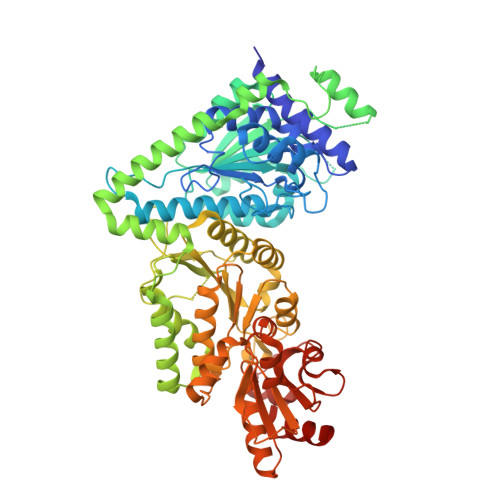 | |
UniProt | |||||
Entity Groups | |||||
| Sequence Clusters | 30% Identity50% Identity70% Identity90% Identity95% Identity100% Identity | ||||
| UniProt Group | Q9I5Y8 | ||||
Sequence AnnotationsExpand | |||||
Reference Sequence | |||||
| Ligands 2 Unique | |||||
|---|---|---|---|---|---|
| ID | Chains | Name / Formula / InChI Key | 2D Diagram | 3D Interactions | |
| EDO Download:Ideal Coordinates CCD File | B [auth A] | 1,2-ETHANEDIOL C2 H6 O2 LYCAIKOWRPUZTN-UHFFFAOYSA-N |  | ||
| CA Download:Ideal Coordinates CCD File | C [auth A], D [auth A] | CALCIUM ION Ca BHPQYMZQTOCNFJ-UHFFFAOYSA-N |  | ||
| Length ( Å ) | Angle ( ˚ ) |
|---|---|
| a = 76.15 | α = 90 |
| b = 76.15 | β = 90 |
| c = 193.61 | γ = 120 |
| Software Name | Purpose |
|---|---|
| XDS | data reduction |
| REFMAC | refinement |
| PDB_EXTRACT | data extraction |
| XSCALE | data scaling |
| BALBES | phasing |


