4WVC

Crystal structure of GH63 mannosylglycerate hydrolase from Thermus thermophilus HB8 in complex with Tris and D-glycerate


Experimental Data Snapshot

  • Method: X-RAY DIFFRACTION
  • Resolution: 2.10 Å
  • R-Value Free: 0.280 
  • R-Value Work: 0.245 
  • R-Value Observed: 0.247 

wwPDB Validation   3D Report Full Report


This is version 1.3 of the entry. See complete history


Literature

Crystal structure and substrate-binding mode of GH63 mannosylglycerate hydrolase from Thermus thermophilus HB8.

Miyazaki, T.Ichikawa, M.Iino, H.Nishikawa, A.Tonozuka, T.

(2015) J Struct Biol 190: 21-30

  • DOI: https://doi.org/10.1016/j.jsb.2015.02.006
  • Primary Citation of Related Structures:  
    4WVA, 4WVB, 4WVC

  • PubMed Abstract: 

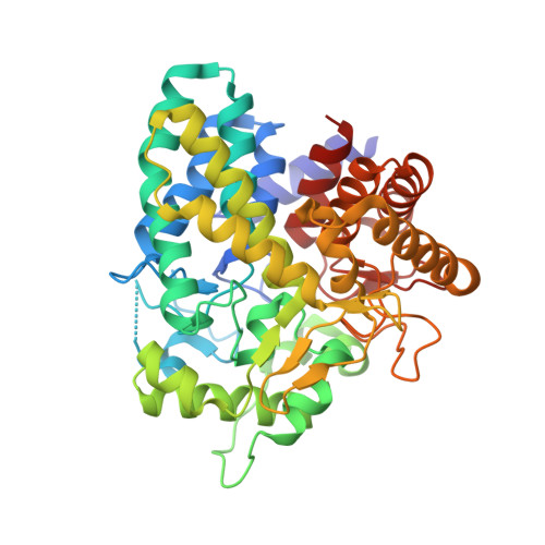
    Glycoside hydrolase family 63 (GH63) proteins are found in eukaryotes such as processing α-glucosidase I and also many bacteria and archaea. Recent studies have identified two bacterial and one plant GH63 mannosylglycerate hydrolases that act on both glucosylglycerate and mannosylglycerate, which are compatible solutes found in many thermophilic prokaryotes and some plants. Here we report the 1.67-Å crystal structure of one of these GH63 mannosylglycerate hydrolases, Tt8MGH from Thermus thermophilus HB8, which is 99% homologous to mannosylglycerate hydrolase from T. thermophilus HB27. Tt8MGH consists of a single (α/α)6-barrel catalytic domain with two additional helices and two long loops which form a homotrimer. The structures of this protein in complexes with glucose or glycerate were also determined at 1.77- or 2.10-Å resolution, respectively. A comparison of these structures revealed that the conformations of three flexible loops were largely different from each other. The conformational changes may be induced by ligand binding and serve to form finger-like structures for holding substrates. These findings represent the first-ever proposed substrate recognition mechanism for GH63 mannosylglycerate hydrolase.


  • Organizational Affiliation

    Department of Applied Biological Science, Tokyo University of Agriculture and Technology, 3-5-8 Saiwai-cho, Fuchu, Tokyo 183-8509, Japan.


Macromolecules
Find similar proteins by:  (by identity cutoff)  |  3D Structure
Entity ID: 1
MoleculeChains Sequence LengthOrganismDetailsImage
Uncharacterized protein
A, B
420Thermus thermophilus HB8Mutation(s): 0 
Gene Names: TTHA0978
UniProt
Find proteins for Q5SJN0 (Thermus thermophilus (strain ATCC 27634 / DSM 579 / HB8))
Explore Q5SJN0 
Go to UniProtKB:  Q5SJN0
Entity Groups  
Sequence Clusters30% Identity50% Identity70% Identity90% Identity95% Identity100% Identity
UniProt GroupQ5SJN0
Sequence Annotations
Expand
  • Reference Sequence
Experimental Data & Validation

Experimental Data

  • Method: X-RAY DIFFRACTION
  • Resolution: 2.10 Å
  • R-Value Free: 0.280 
  • R-Value Work: 0.245 
  • R-Value Observed: 0.247 
  • Space Group: P 3 2 1
Unit Cell:
Length ( Å )Angle ( ˚ )
a = 92.94α = 90
b = 92.94β = 90
c = 190.401γ = 120
Software Package:
Software NamePurpose
REFMACrefinement
MOSFLMdata processing
SCALAdata scaling
MOLREPphasing
Cootmodel building
SCALEPACKdata reduction

Structure Validation

View Full Validation Report


Entry History 

Deposition Data

Revision History  (Full details and data files)

  • Version 1.0: 2015-03-11
    Type: Initial release
  • Version 1.1: 2015-04-22
    Changes: Database references
  • Version 1.2: 2020-02-05
    Changes: Data collection, Database references, Derived calculations, Source and taxonomy
  • Version 1.3: 2024-03-20
    Changes: Data collection, Database references