


Crystal structure of UDP-glucose 4-epimerase from Brucella ovis in complex with NAD
SSGCID, Seattle Structural Genomics Center for Infectious Disease (SSGCID)To be published.
Experimental Data Snapshot
Starting Model: experimental
View more details
Entity ID: 1 | |||||
|---|---|---|---|---|---|
| Molecule | Chains | Sequence Length | Organism | Details | Image |
| UDP-glucose 4-epimerase | 336 | Brucella ovis ATCC 25840 | Mutation(s): 2 Gene Names: galE-1, BOV_A0474 EC: 5.1.3.2 | 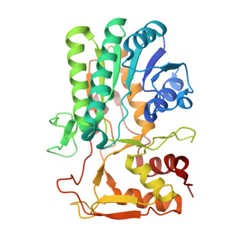 | |
UniProt | |||||
Find proteins for A0A0H3ASR6 (Brucella ovis (strain ATCC 25840 / 63/290 / NCTC 10512)) Explore A0A0H3ASR6 Go to UniProtKB: A0A0H3ASR6 | |||||
Entity Groups | |||||
| Sequence Clusters | 30% Identity50% Identity70% Identity90% Identity95% Identity100% Identity | ||||
| UniProt Group | A0A0H3ASR6 | ||||
Sequence AnnotationsExpand | |||||
Reference Sequence | |||||
| Ligands 3 Unique | |||||
|---|---|---|---|---|---|
| ID | Chains | Name / Formula / InChI Key | 2D Diagram | 3D Interactions | |
| NAD Download:Ideal Coordinates CCD File | B [auth A] | NICOTINAMIDE-ADENINE-DINUCLEOTIDE C21 H27 N7 O14 P2 BAWFJGJZGIEFAR-NNYOXOHSSA-N |  | ||
| PO4 Download:Ideal Coordinates CCD File | D [auth A] | PHOSPHATE ION O4 P NBIIXXVUZAFLBC-UHFFFAOYSA-K |  | ||
| ZN Download:Ideal Coordinates CCD File | C [auth A] | ZINC ION Zn PTFCDOFLOPIGGS-UHFFFAOYSA-N |  | ||
| Length ( Å ) | Angle ( ˚ ) |
|---|---|
| a = 76.48 | α = 90 |
| b = 86.46 | β = 90 |
| c = 126.85 | γ = 90 |
| Software Name | Purpose |
|---|---|
| XDS | data reduction |
| XSCALE | data scaling |
| PHASER | phasing |
| ARP | model building |
| Coot | model building |
| PHENIX | refinement |
| PDB_EXTRACT | data extraction |


