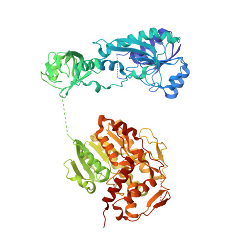
4WKG

The crystal structure of apo ArnA features an unexpected central binding pocket and provides an explanation for enzymatic coop-erativity


Experimental Data Snapshot

  • Method: X-RAY DIFFRACTION
  • Resolution: 2.70 Å
  • R-Value Free: 0.232 
  • R-Value Work: 0.205 
  • R-Value Observed: 0.207 

Starting Model: experimental
View more details

wwPDB Validation   3D Report Full Report


This is version 1.3 of the entry. See complete history


Literature

The structure of apo ArnA features an unexpected central binding pocket and provides an explanation for enzymatic cooperativity.

Fischer, U.Hertlein, S.Grimm, C.

(2015) Acta Crystallogr D Biol Crystallogr 71: 687-696

  • DOI: https://doi.org/10.1107/S1399004714026686
  • Primary Citation of Related Structures:  
    4WKG

  • PubMed Abstract: 

    The bacterial protein ArnA is an essential enzyme in the pathway leading to the modification of lipid A with the pentose sugar 4-amino-4-deoxy-L-arabinose. This modification confers resistance to polymyxins, which are antibiotics that are used as a last resort to treat infections with multiple drug-resistant Gram-negative bacteria. ArnA contains two domains with distinct catalytic functions: a dehydrogenase domain and a transformylase domain. The protein forms homohexamers organized as a dimer of trimers. Here, the crystal structure of apo ArnA is presented and compared with its ATP- and UDP-glucuronic acid-bound counterparts. The comparison reveals major structural rearrangements in the dehydrogenase domain that lead to the formation of a previously unobserved binding pocket at the centre of each ArnA trimer in its apo state. In the crystal structure, this pocket is occupied by a DTT molecule. It is shown that formation of the pocket is linked to a cascade of structural rearrangements that emerge from the NAD(+)-binding site. Based on these findings, a small effector molecule is postulated that binds to the central pocket and modulates the catalytic properties of ArnA. Furthermore, the discovered conformational changes provide a mechanistic explanation for the strong cooperative effect recently reported for the ArnA dehydrogenase function.


  • Organizational Affiliation

    Department of Biochemistry, Biocenter of the University of Würzburg, Am Hubland, 97074 Würzburg, Germany.


Macromolecules
Find similar proteins by:  (by identity cutoff)  |  3D Structure
Entity ID: 1
MoleculeChains Sequence LengthOrganismDetailsImage
Bifunctional polymyxin resistance protein ArnA
A, B, C, D, E
A, B, C, D, E, F
660Escherichia coli K-12Mutation(s): 0 
EC: 2.1.2.13 (PDB Primary Data), 1.1.1.305 (PDB Primary Data)
UniProt
Find proteins for P77398 (Escherichia coli (strain K12))
Explore P77398 
Go to UniProtKB:  P77398
Entity Groups  
Sequence Clusters30% Identity50% Identity70% Identity90% Identity95% Identity100% Identity
UniProt GroupP77398
Sequence Annotations
Expand
  • Reference Sequence
Experimental Data & Validation

Experimental Data

  • Method: X-RAY DIFFRACTION
  • Resolution: 2.70 Å
  • R-Value Free: 0.232 
  • R-Value Work: 0.205 
  • R-Value Observed: 0.207 
  • Space Group: P 1
Unit Cell:
Length ( Å )Angle ( ˚ )
a = 106.72α = 81.92
b = 112.82β = 82.96
c = 113.61γ = 83.8
Software Package:
Software NamePurpose
PHENIXrefinement
XDSdata reduction
XSCALEdata scaling
PHASERphasing

Structure Validation

View Full Validation Report


Entry History 

Deposition Data

  • Released Date: 2014-12-17 
  • Deposition Author(s): Grimm, C.

Revision History  (Full details and data files)

  • Version 1.0: 2014-12-17
    Type: Initial release
  • Version 1.1: 2015-03-11
    Changes: Database references
  • Version 1.2: 2016-07-20
    Changes: Database references
  • Version 1.3: 2024-01-10
    Changes: Data collection, Database references, Refinement description