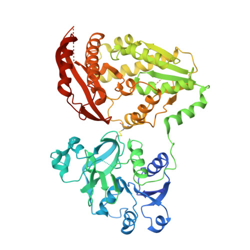
4WIO

Crystal structure of the C89A GMP synthetase inactive mutant from Plasmodium falciparum in complex with glutamine


Experimental Data Snapshot

  • Method: X-RAY DIFFRACTION
  • Resolution: 3.15 Å
  • R-Value Free: 0.306 
  • R-Value Work: 0.263 
  • R-Value Observed: 0.265 

Starting Model: experimental
View more details

wwPDB Validation   3D Report Full Report


This is version 1.2 of the entry. See complete history


Literature

Active site coupling in Plasmodium falciparum GMP synthetase is triggered by domain rotation.

Ballut, L.Violot, S.Shivakumaraswamy, S.Thota, L.P.Sathya, M.Kunala, J.Dijkstra, B.W.Terreux, R.Haser, R.Balaram, H.Aghajari, N.

(2015) Nat Commun 6: 8930-8930

  • DOI: https://doi.org/10.1038/ncomms9930
  • Primary Citation of Related Structures:  
    4WIM, 4WIN, 4WIO

  • PubMed Abstract: 

    GMP synthetase (GMPS), a key enzyme in the purine biosynthetic pathway performs catalysis through a coordinated process across two catalytic pockets for which the mechanism remains unclear. Crystal structures of Plasmodium falciparum GMPS in conjunction with mutational and enzyme kinetic studies reported here provide evidence that an 85° rotation of the GATase domain is required for ammonia channelling and thus for the catalytic activity of this two-domain enzyme. We suggest that conformational changes in helix 371-375 holding catalytic residues and in loop 376-401 along the rotation trajectory trigger the different steps of catalysis, and establish the central role of Glu374 in allostery and inter-domain crosstalk. These studies reveal the mechanism of domain rotation and inter-domain communication, providing a molecular framework for the function of all single polypeptide GMPSs and form a solid basis for rational drug design targeting this therapeutically important enzyme.


  • Organizational Affiliation

    BioCrystallography and Structural Biology of Therapeutic Targets Group, Molecular and Structural Bases of Infectious Systems, UMR5086 CNRS-University of Lyon 1, 7 passage du Vercors, 69367 Lyon Cedex 07, France.


Macromolecules
Find similar proteins by:  (by identity cutoff)  |  3D Structure
Entity ID: 1
MoleculeChains Sequence LengthOrganismDetailsImage
GMP synthetase567Plasmodium falciparum 3D7Mutation(s): 1 
Gene Names: PF10_0123
EC: 6.3.5.2
UniProt
Find proteins for Q8IJR9 (Plasmodium falciparum (isolate 3D7))
Explore Q8IJR9 
Go to UniProtKB:  Q8IJR9
Entity Groups  
Sequence Clusters30% Identity50% Identity70% Identity90% Identity95% Identity100% Identity
UniProt GroupQ8IJR9
Sequence Annotations
Expand
  • Reference Sequence
Small Molecules
Ligands 1 Unique
IDChains Name / Formula / InChI Key2D Diagram3D Interactions
GLN
Query on GLN

Download Ideal Coordinates CCD File 
B [auth A]GLUTAMINE
C5 H10 N2 O3
ZDXPYRJPNDTMRX-VKHMYHEASA-N
Experimental Data & Validation

Experimental Data

  • Method: X-RAY DIFFRACTION
  • Resolution: 3.15 Å
  • R-Value Free: 0.306 
  • R-Value Work: 0.263 
  • R-Value Observed: 0.265 
  • Space Group: P 43 21 2
Unit Cell:
Length ( Å )Angle ( ˚ )
a = 76.92α = 90
b = 76.92β = 90
c = 204.78γ = 90
Software Package:
Software NamePurpose
PHENIXrefinement
XDSdata reduction
XSCALEdata scaling
PHASERphasing

Structure Validation

View Full Validation Report


Entry History 

Deposition Data

Revision History  (Full details and data files)

  • Version 1.0: 2015-12-02
    Type: Initial release
  • Version 1.1: 2015-12-09
    Changes: Database references
  • Version 1.2: 2024-01-10
    Changes: Data collection, Database references, Refinement description