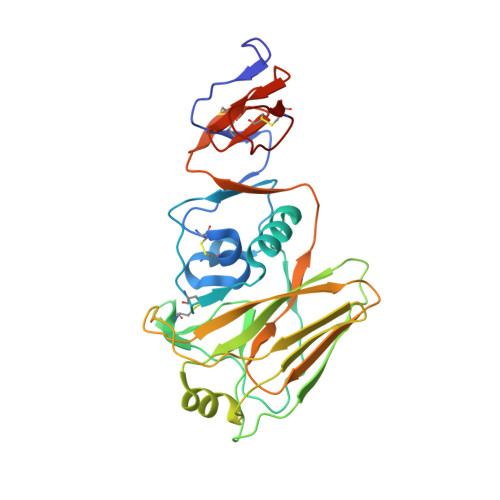
4WE6

The crystal structure of hemagglutinin HA1 domain from influenza virus A/Perth/142/2007(H3N2)


Experimental Data Snapshot

  • Method: X-RAY DIFFRACTION
  • Resolution: 1.90 Å
  • R-Value Free: 0.201 
  • R-Value Work: 0.170 
  • R-Value Observed: 0.171 

wwPDB Validation   3D Report Full Report


Ligand Structure Quality Assessment 


This is version 1.3 of the entry. See complete history


Literature

Structure and receptor binding preferences of recombinant human A(H3N2) virus hemagglutinins.

Yang, H.Carney, P.J.Chang, J.C.Guo, Z.Villanueva, J.M.Stevens, J.

(2015) Virology 477C: 18-31

  • DOI: https://doi.org/10.1016/j.virol.2014.12.024
  • Primary Citation of Related Structures:  
    4WE4, 4WE5, 4WE6, 4WE7, 4WE8, 4WE9, 4WEA

  • PubMed Abstract: 

    A(H3N2) influenza viruses have circulated in humans since 1968, and antigenic drift of the hemagglutinin (HA) protein continues to be a driving force that allows the virus to escape the human immune response. Since the major antigenic sites of the HA overlap into the receptor binding site (RBS) of the molecule, the virus constantly struggles to effectively adapt to host immune responses, without compromising its functionality. Here, we have structurally assessed the evolution of the A(H3N2) virus HA RBS, using an established recombinant expression system. Glycan binding specificities of nineteen A(H3N2) influenza virus HAs, each a component of the seasonal influenza vaccine between 1968 and 2012, were analyzed. Results suggest that while its receptor-binding site has evolved from one that can bind a broad range of human receptor analogs to one with a more restricted binding profile for longer glycans, the virus continues to circulate and transmit efficiently among humans.


  • Organizational Affiliation

    Influenza Division, National Center for Immunization and Respiratory Diseases, Centers for Disease Control and Prevention, Atlanta, GA 30333, USA.


Macromolecules
Find similar proteins by:  (by identity cutoff)  |  3D Structure
Entity ID: 1
MoleculeChains Sequence LengthOrganismDetailsImage
Hemagglutinin HA1 chain
A, B
279Influenza A virus (A/Perth/142/2007(H3N2))Mutation(s): 0 
Gene Names: HA
UniProt
Find proteins for B2VNP1 (Influenza A virus)
Explore B2VNP1 
Go to UniProtKB:  B2VNP1
Entity Groups  
Sequence Clusters30% Identity50% Identity70% Identity90% Identity95% Identity100% Identity
UniProt GroupB2VNP1
Glycosylation
Glycosylation Sites: 2
Sequence Annotations
Expand
  • Reference Sequence
Small Molecules
Ligands 2 Unique
IDChains Name / Formula / InChI Key2D Diagram3D Interactions
NAG
Query on NAG

Download Ideal Coordinates CCD File 
C [auth A],
D [auth A],
G [auth B],
H [auth B]
2-acetamido-2-deoxy-beta-D-glucopyranose
C8 H15 N O6
OVRNDRQMDRJTHS-FMDGEEDCSA-N
GOL
Query on GOL

Download Ideal Coordinates CCD File 
E [auth A],
F [auth A],
I [auth B],
J [auth B]
GLYCEROL
C3 H8 O3
PEDCQBHIVMGVHV-UHFFFAOYSA-N
Experimental Data & Validation

Experimental Data

  • Method: X-RAY DIFFRACTION
  • Resolution: 1.90 Å
  • R-Value Free: 0.201 
  • R-Value Work: 0.170 
  • R-Value Observed: 0.171 
  • Space Group: P 1 2 1
Unit Cell:
Length ( Å )Angle ( ˚ )
a = 42.472α = 90
b = 50.567β = 90.08
c = 182.256γ = 90
Software Package:
Software NamePurpose
PHENIXrefinement

Structure Validation

View Full Validation Report


Ligand Structure Quality Assessment 


Entry History 

Deposition Data

Revision History  (Full details and data files)

  • Version 1.0: 2015-02-11
    Type: Initial release
  • Version 1.1: 2017-09-27
    Changes: Data collection, Database references, Derived calculations, Other, Source and taxonomy, Structure summary
  • Version 1.2: 2020-07-29
    Type: Remediation
    Reason: Carbohydrate remediation
    Changes: Data collection, Derived calculations, Refinement description, Structure summary
  • Version 1.3: 2023-12-27
    Changes: Data collection, Database references, Structure summary