4WAC | pdb_00004wac

Crystal Structure of TarM


Experimental Data Snapshot

  • Method: X-RAY DIFFRACTION
  • Resolution: 2.40 Å
  • R-Value Free: 
    0.252 (Depositor), 0.259 (DCC) 
  • R-Value Work: 
    0.215 (Depositor), 0.224 (DCC) 
  • R-Value Observed: 
    0.217 (Depositor) 

wwPDB Validation 3D Report Full Report

Validation slider image for 4WAC

This is version 2.1 of the entry. See complete history

Literature

Structural and Enzymatic Analysis of TarM Glycosyltransferase from Staphylococcus aureus Reveals an Oligomeric Protein Specific for the Glycosylation of Wall Teichoic Acid.

Koc, C.Gerlach, D.Beck, S.Peschel, A.Xia, G.Stehle, T.

(2015) J Biological Chem 290: 9874-9885

  • DOI: https://doi.org/10.1074/jbc.M114.619924
  • Primary Citation Related Structures: 
    4WAC, 4WAD

  • PubMed Abstract: 

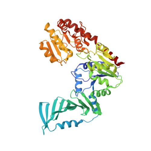
    Anionic glycopolymers known as wall teichoic acids (WTAs) functionalize the peptidoglycan layers of many Gram-positive bacteria. WTAs play central roles in many fundamental aspects of bacterial physiology, and they are important determinants of pathogenesis and antibiotic resistance. A number of enzymes that glycosylate WTA in Staphylococcus aureus have recently been identified. Among these is the glycosyltransferase TarM, a component of the WTA de novo biosynthesis pathway. TarM performs the synthesis of α-O-N-acetylglycosylated poly-5'-phosphoribitol in the WTA structure. We have solved the crystal structure of TarM at 2.4 Å resolution, and we have also determined a structure of the enzyme in complex with its substrate UDP-GlcNAc at 2.8 Å resolution. The protein assembles into a propeller-like homotrimer in which each blade contains a GT-B-type glycosyltransferase domain with a typical Rossmann fold. The enzymatic reaction retains the stereochemistry of the anomeric center of the transferred GlcNAc-moiety on the polyribitol backbone. TarM assembles into a trimer using a novel trimerization domain, here termed the HUB domain. Structure-guided mutagenesis experiments of TarM identify residues critical for enzyme activity, assign a putative role for the HUB in TarM function, and allow us to propose a likely reaction mechanism.


  • Organizational Affiliation
    • From the Interfaculty Institute of Biochemistry, University of Tübingen, 72076 Tübingen, Germany.

Macromolecule Content 

  • Total Structure Weight: 58.35 kDa 
  • Atom Count: 4,194 
  • Modeled Residue Count: 498 
  • Deposited Residue Count: 498 
  • Unique protein chains: 1

Macromolecules

Find similar proteins by:|  3D Structure
Entity ID: 1
MoleculeChains  Sequence LengthOrganismDetailsImage
Glycosyl transferase, group 1 family protein498Staphylococcus aureusMutation(s): 0 
EC: 2.4.1.5 (PDB Primary Data), 2.4.1.52 (PDB Primary Data), 2.4 (UniProt)
UniProt
Find proteins for A0A0D6HUA0 (Staphylococcus aureus)
Explore A0A0D6HUA0 
Go to UniProtKB:  A0A0D6HUA0
Entity Groups
Sequence Clusters30% Identity50% Identity70% Identity90% Identity95% Identity100% Identity
UniProt GroupA0A0D6HUA0
Sequence Annotations
Expand
Reference Sequence

Small Molecules

Ligands 4 Unique
IDChains Name / Formula / InChI Key2D Diagram3D Interactions
PEG

Query on PEG


Download:Ideal Coordinates CCD File
D [auth A]DI(HYDROXYETHYL)ETHER
C4 H10 O3
MTHSVFCYNBDYFN-UHFFFAOYSA-N
IMD

Query on IMD


Download:Ideal Coordinates CCD File
B [auth A]IMIDAZOLE
C3 H5 N2
RAXXELZNTBOGNW-UHFFFAOYSA-O
EDO

Query on EDO


Download:Ideal Coordinates CCD File
E [auth A],
G [auth A]
1,2-ETHANEDIOL
C2 H6 O2
LYCAIKOWRPUZTN-UHFFFAOYSA-N
ACT

Query on ACT


Download:Ideal Coordinates CCD File
C [auth A],
F [auth A]
ACETATE ION
C2 H3 O2
QTBSBXVTEAMEQO-UHFFFAOYSA-M

Experimental Data & Validation

Experimental Data

  • Method: X-RAY DIFFRACTION
  • Resolution: 2.40 Å
  • R-Value Free:  0.252 (Depositor), 0.259 (DCC) 
  • R-Value Work:  0.215 (Depositor), 0.224 (DCC) 
  • R-Value Observed: 0.217 (Depositor) 
Space Group: P 63 2 2
Unit Cell:
Length ( Å )Angle ( ˚ )
a = 124.749α = 90
b = 124.749β = 90
c = 223.255γ = 120
Software Package:
Software NamePurpose
XDSdata reduction
XSCALEdata scaling
SHARPphasing
Cootmodel building
PHENIXrefinement
PDB_EXTRACTdata extraction

Structure Validation

View Full Validation Report


Entry History 

& Funding Information

Deposition Data


Funding OrganizationLocationGrant Number
German Research FoundationGermany--

Revision History  (Full details and data files)

  • Version 1.0: 2015-02-25
    Type: Initial release
  • Version 1.1: 2015-03-04
    Changes: Derived calculations
  • Version 1.2: 2015-04-22
    Changes: Database references
  • Version 2.0: 2017-09-06
    Changes: Advisory, Atomic model, Author supporting evidence, Derived calculations, Refinement description
  • Version 2.1: 2024-05-08
    Changes: Data collection, Database references