


Crystal structure of XacCel5A in the native form
Paiva, J.H., Murakami, M.T.To be published.
Experimental Data Snapshot
Starting Model: experimental
View more details
wwPDB Validation 3D Report Full Report

Entity ID: 1 | |||||
|---|---|---|---|---|---|
| Molecule | Chains | Sequence Length | Organism | Details | Image |
| Cellulase | 332 | Xanthomonas citri subsp. citri 306 | Mutation(s): 0 Gene Names: egl, XAC0030 | 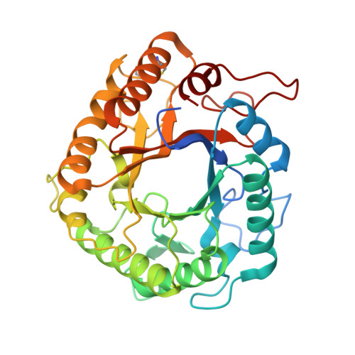 | |
| Ligands 1 Unique | |||||
|---|---|---|---|---|---|
| ID | Chains | Name / Formula / InChI Key | 2D Diagram | 3D Interactions | |
| CAC Download:Ideal Coordinates CCD File | B [auth A], C [auth A] | CACODYLATE ION C2 H6 As O2 OGGXGZAMXPVRFZ-UHFFFAOYSA-M |  | ||
| Length ( Å ) | Angle ( ˚ ) |
|---|---|
| a = 79.367 | α = 90 |
| b = 81.73 | β = 90 |
| c = 48.232 | γ = 90 |
| Software Name | Purpose |
|---|---|
| XDS | data scaling |
| REFMAC | refinement |
| PDB_EXTRACT | data extraction |
| XSCALE | data scaling |
| Funding Organization | Location | Grant Number |
|---|---|---|
| Sao Paulo Research Foundation (FAPESP) | Brazil | 2013/13309-0 and 2014/07135-1 |


