4V7G | pdb_00004v7g

Crystal Structure of Lumazine Synthase from Bacillus Anthracis


Experimental Data Snapshot

  • Method: X-RAY DIFFRACTION
  • Resolution: 3.50 Å
  • R-Value Free: 
    0.286 (Depositor), 0.355 (DCC) 
  • R-Value Work: 
    0.235 (Depositor), 0.351 (DCC) 
  • R-Value Observed: 
    0.236 (Depositor) 

Starting Model: experimental
View more details

wwPDB Validation 3D Report Full Report

Validation slider image for 4V7G

This is version 1.2 of the entry. See complete history

Literature

Structural study and thermodynamic characterization of inhibitor binding to lumazine synthase from Bacillus anthracis.

Morgunova, E.Illarionov, B.Saller, S.Popov, A.Sambaiah, T.Bacher, A.Cushman, M.Fischer, M.Ladenstein, R.

(2010) Acta Crystallogr D Biol Crystallogr 66: 1001-1011

  • DOI: https://doi.org/10.1107/S0907444910029690
  • Primary Citation Related Structures: 
    4V7G

  • PubMed Abstract: 

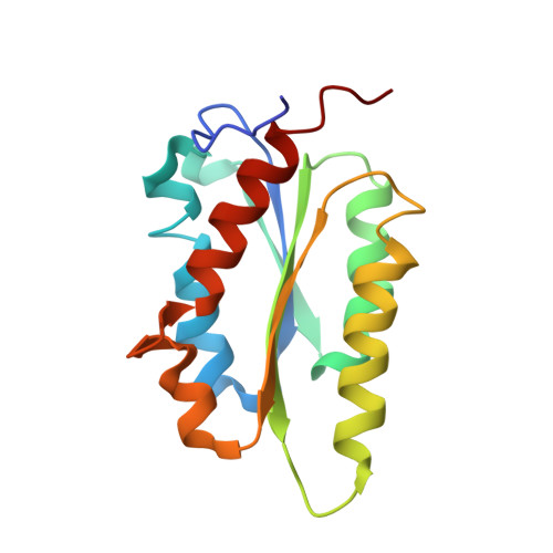
    The crystal structure of lumazine synthase from Bacillus anthracis was solved by molecular replacement and refined to R(cryst) = 23.7% (R(free) = 28.4%) at a resolution of 3.5 A. The structure reveals the icosahedral symmetry of the enzyme and specific features of the active site that are unique in comparison with previously determined orthologues. The application of isothermal titration calorimetry in combination with enzyme kinetics showed that three designed pyrimidine derivatives bind to lumazine synthase with micromolar dissociation constants and competitively inhibit the catalytic reaction. Structure-based modelling suggested the binding modes of the inhibitors in the active site and allowed an estimation of the possible contacts formed upon binding. The results provide a structural framework for the design of antibiotics active against B. anthracis.


  • Organizational Affiliation
    • Karolinska Institutet NOVUM, Center of Structural Biochemistry, Huddinge, Sweden.

Macromolecule Content 

  • Total Structure Weight: 1,473.18 kDa 
  • Atom Count: 102,534 
  • Modeled Residue Count: 13,680 
  • Deposited Residue Count: 13,770 
  • Unique protein chains: 1

Macromolecules

Find similar proteins by:|  3D Structure
Entity ID: 1
MoleculeChains  Sequence LengthOrganismDetailsImage
6,7-dimethyl-8-ribityllumazine synthase153Bacillus anthracisMutation(s): 0 
Gene Names: ribH
EC: 2.5.1.78
UniProt
Find proteins for Q81MB5 (Bacillus anthracis)
Explore Q81MB5 
Go to UniProtKB:  Q81MB5
Entity Groups
Sequence Clusters30% Identity50% Identity70% Identity90% Identity95% Identity100% Identity
UniProt GroupQ81MB5
Sequence Annotations
Expand
Reference Sequence

Small Molecules

Ligands 1 Unique
IDChains Name / Formula / InChI Key2D Diagram3D Interactions
PO4

Query on PO4


Download:Ideal Coordinates CCD File
AD [auth AO]
AE [auth BK]
AF [auth CG]
BD [auth AP]
BE [auth BL]
AD [auth AO],
AE [auth BK],
AF [auth CG],
BD [auth AP],
BE [auth BL],
BF [auth CH],
CD [auth AQ],
CE [auth BM],
CF [auth CI],
DD [auth AR],
DE [auth BN],
DF [auth CJ],
ED [auth AS],
EE [auth BO],
EF [auth CK],
FD [auth AT],
FE [auth BP],
FF [auth CL],
GD [auth AU],
GE [auth BQ],
GF [auth CM],
HD [auth AV],
HE [auth BR],
HF [auth CN],
ID [auth AW],
IE [auth BS],
IF [auth CO],
JD [auth AX],
JE [auth BT],
JF [auth CP],
KD [auth AY],
KE [auth BU],
KF [auth CQ],
LD [auth AZ],
LE [auth BV],
LF [auth CR],
MC [auth AA],
MD [auth A1],
ME [auth BW],
MF [auth CS],
NC [auth AB],
ND [auth A2],
NE [auth BX],
NF [auth CT],
OC [auth AC],
OD [auth A3],
OE [auth BY],
OF [auth CU],
PC [auth AD],
PD [auth A4],
PE [auth BZ],
PF [auth CV],
QC [auth AE],
QD [auth BA],
QE [auth B1],
QF [auth CW],
RC [auth AF],
RD [auth BB],
RE [auth B2],
RF [auth CX],
SC [auth AG],
SD [auth BC],
SE [auth B3],
SF [auth CY],
TC [auth AH],
TD [auth BD],
TE [auth B4],
TF [auth CZ],
UC [auth AI],
UD [auth BE],
UE [auth CA],
UF [auth C1],
VC [auth AJ],
VD [auth BF],
VE [auth CB],
VF [auth C2],
WC [auth AK],
WD [auth BG],
WE [auth CC],
WF [auth C3],
XC [auth AL],
XD [auth BH],
XE [auth CD],
XF [auth C4],
YC [auth AM],
YD [auth BI],
YE [auth CE],
ZC [auth AN],
ZD [auth BJ],
ZE [auth CF]
PHOSPHATE ION
O4 P
NBIIXXVUZAFLBC-UHFFFAOYSA-K

Experimental Data & Validation

Experimental Data

  • Method: X-RAY DIFFRACTION
  • Resolution: 3.50 Å
  • R-Value Free:  0.286 (Depositor), 0.355 (DCC) 
  • R-Value Work:  0.235 (Depositor), 0.351 (DCC) 
  • R-Value Observed: 0.236 (Depositor) 
Space Group: P 21 21 2
Unit Cell:
Length ( Å )Angle ( ˚ )
a = 157.201α = 90
b = 222.237β = 90
c = 473.488γ = 90
Software Package:
Software NamePurpose
ADSCdata collection
MOLREPphasing
FFFEARmodel building
PHENIXrefinement
XDSdata reduction
SCALAdata scaling
FFFEARphasing

Structure Validation

View Full Validation Report


Entry History 

Deposition Data

Revision History  (Full details and data files)

  • Version 1.0: 2014-07-09
    Type: Initial release
  • Version 1.1: 2014-12-10
    Changes: Other
  • Version 1.2: 2023-09-20
    Changes: Data collection, Database references, Refinement description