4V1X

The structure of the hexameric atrazine chlorohydrolase, AtzA


Experimental Data Snapshot

  • Method: X-RAY DIFFRACTION
  • Resolution: 2.20 Å
  • R-Value Free: 0.196 
  • R-Value Work: 0.166 
  • R-Value Observed: 0.167 

wwPDB Validation   3D Report Full Report


This is version 1.1 of the entry. See complete history


Literature

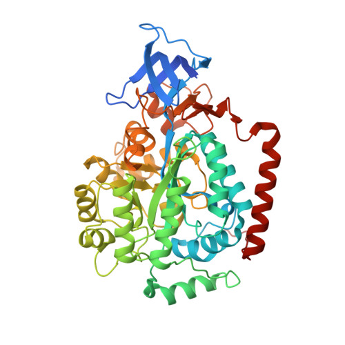
The Structure of the Hexameric Atrazine Chlorohydrolase Atza.

Peat, T.S.Newman, J.Balotra, S.Lucent, D.Warden, A.C.Scott, C.

(2015) Acta Crystallogr D Biol Crystallogr 71: 710

  • DOI: https://doi.org/10.1107/S1399004715000619
  • Primary Citation of Related Structures:  
    4V1X, 4V1Y

  • PubMed Abstract: 

    Atrazine chlorohydrolase (AtzA) was discovered and purified in the early 1990s from soil that had been exposed to the widely used herbicide atrazine. It was subsequently found that this enzyme catalyzes the first and necessary step in the breakdown of atrazine by the soil organism Pseudomonas sp. strain ADP. Although it has taken 20 years, a crystal structure of the full hexameric form of AtzA has now been obtained. AtzA is less well adapted to its physiological role (i.e. atrazine dechlorination) than the alternative metal-dependent atrazine chlorohydrolase (TrzN), with a substrate-binding pocket that is under considerable strain and for which the substrate is a poor fit.


  • Organizational Affiliation

    CSIRO Biomedical Manufacturing, Parkville, Australia.


Macromolecules
Find similar proteins by:  (by identity cutoff)  |  3D Structure
Entity ID: 1
MoleculeChains Sequence LengthOrganismDetailsImage
ATRAZINE CHLOROHYDROLASE
A, B, C, D, E
A, B, C, D, E, F
494Pseudomonas sp. ADPMutation(s): 0 
EC: 3.8.1.8
UniProt
Find proteins for P72156 (Pseudomonas sp. (strain ADP))
Explore P72156 
Go to UniProtKB:  P72156
Entity Groups  
Sequence Clusters30% Identity50% Identity70% Identity90% Identity95% Identity100% Identity
UniProt GroupP72156
Sequence Annotations
Expand
  • Reference Sequence
Small Molecules
Ligands 2 Unique
IDChains Name / Formula / InChI Key2D Diagram3D Interactions
PEG
Query on PEG

Download Ideal Coordinates CCD File 
H [auth A]
J [auth B]
L [auth C]
N [auth D]
P [auth E]
H [auth A],
J [auth B],
L [auth C],
N [auth D],
P [auth E],
R [auth F]
DI(HYDROXYETHYL)ETHER
C4 H10 O3
MTHSVFCYNBDYFN-UHFFFAOYSA-N
FE
Query on FE

Download Ideal Coordinates CCD File 
G [auth A]
I [auth B]
K [auth C]
M [auth D]
O [auth E]
G [auth A],
I [auth B],
K [auth C],
M [auth D],
O [auth E],
Q [auth F]
FE (III) ION
Fe
VTLYFUHAOXGGBS-UHFFFAOYSA-N
Experimental Data & Validation

Experimental Data

  • Method: X-RAY DIFFRACTION
  • Resolution: 2.20 Å
  • R-Value Free: 0.196 
  • R-Value Work: 0.166 
  • R-Value Observed: 0.167 
  • Space Group: P 21 21 21
Unit Cell:
Length ( Å )Angle ( ˚ )
a = 117.269α = 90
b = 146.056β = 90
c = 196.361γ = 90
Software Package:
Software NamePurpose
REFMACrefinement
XDSdata reduction
Aimlessdata scaling
PHASERphasing

Structure Validation

View Full Validation Report


Entry History 

Deposition Data

Revision History  (Full details and data files)

  • Version 1.0: 2015-03-11
    Type: Initial release
  • Version 1.1: 2015-03-25
    Changes: Database references