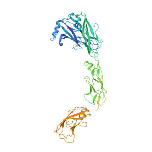
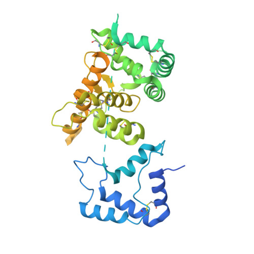
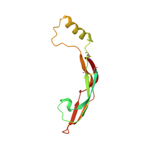
4UX8

RET recognition of GDNF-GFRalpha1 ligand by a composite binding site promotes membrane-proximal self-association


Experimental Data Snapshot

  • Method: ELECTRON MICROSCOPY
  • Resolution: 24.0 Å
  • Aggregation State: PARTICLE 
  • Reconstruction Method: SINGLE PARTICLE 

wwPDB Validation   3D Report Full Report


This is version 2.0 of the entry. See complete history


Literature

Ret Recognition of Gdnf-Gfralpha1 Ligand by a Composite Binding Site Promotes Membrane-Proximal Self-Association.

Goodman, K.M.Kjaer, S.Beuron, F.Knowles, P.P.Nawrotek, A.Burns, E.M.Purkiss, A.G.George, R.Santoro, M.Morris, E.P.Mcdonald, N.Q.

(2014) Cell Rep 8: 1894

  • DOI: https://doi.org/10.1016/j.celrep.2014.08.040
  • Primary Citation of Related Structures:  
    4UX8

  • PubMed Abstract: 

    The RET receptor tyrosine kinase is essential to vertebrate development and implicated in multiple human diseases. RET binds a cell surface bipartite ligand comprising a GDNF family ligand and a GFRα coreceptor, resulting in RET transmembrane signaling. We present a hybrid structural model, derived from electron microscopy (EM) and low-angle X-ray scattering (SAXS) data, of the RET extracellular domain (RET(ECD)), GDNF, and GFRα1 ternary complex, defining the basis for ligand recognition. RET(ECD) envelopes the dimeric ligand complex through a composite binding site comprising four discrete contact sites. The GFRα1-mediated contacts are crucial, particularly close to the invariant RET calcium-binding site, whereas few direct contacts are made by GDNF, explaining how distinct ligand/coreceptor pairs are accommodated. The RET(ECD) cysteine-rich domain (CRD) contacts both ligand components and makes homotypic membrane-proximal interactions occluding three different antibody epitopes. Coupling of these CRD-mediated interactions suggests models for ligand-induced RET activation and ligand-independent oncogenic deregulation.


  • Organizational Affiliation

    Structural Biology Laboratory, Cancer Research UK, London Research Institute, 44 Lincoln's Inn Fields, London WC2A 3LY, UK.


Macromolecules
Find similar proteins by:  (by identity cutoff)  |  3D Structure
Entity ID: 1
MoleculeChains Sequence LengthOrganismDetailsImage
PROTO-ONCOGENE TYROSINE-PROTEIN KINASE RECEPTOR RET
A, B
607Homo sapiensMutation(s): 2 
EC: 2.7.10.1
UniProt & NIH Common Fund Data Resources
Find proteins for P07949 (Homo sapiens)
Explore P07949 
Go to UniProtKB:  P07949
PHAROS:  P07949
GTEx:  ENSG00000165731 
Entity Groups  
Sequence Clusters30% Identity50% Identity70% Identity90% Identity95% Identity100% Identity
UniProt GroupP07949
Sequence Annotations
Expand
  • Reference Sequence
Find similar proteins by:  (by identity cutoff)  |  3D Structure
Entity ID: 2
MoleculeChains Sequence LengthOrganismDetailsImage
GDNF FAMILY RECEPTOR ALPHA-1
C, E
463Rattus norvegicusMutation(s): 0 
UniProt
Find proteins for Q62997 (Rattus norvegicus)
Explore Q62997 
Go to UniProtKB:  Q62997
Entity Groups  
Sequence Clusters30% Identity50% Identity70% Identity90% Identity95% Identity100% Identity
UniProt GroupQ62997
Sequence Annotations
Expand
  • Reference Sequence
Find similar proteins by:  (by identity cutoff)  |  3D Structure
Entity ID: 3
MoleculeChains Sequence LengthOrganismDetailsImage
GLIAL CELL LINE-DERIVED NEUROTROPHIC FACTOR
D, F
134Homo sapiensMutation(s): 0 
UniProt & NIH Common Fund Data Resources
Find proteins for P39905 (Homo sapiens)
Explore P39905 
Go to UniProtKB:  P39905
PHAROS:  P39905
GTEx:  ENSG00000168621 
Entity Groups  
Sequence Clusters30% Identity50% Identity70% Identity90% Identity95% Identity100% Identity
UniProt GroupP39905
Glycosylation
Glycosylation Sites: 1Go to GlyGen: P39905-1
Sequence Annotations
Expand
  • Reference Sequence
Oligosaccharides

Help

Entity ID: 4
MoleculeChains Length2D Diagram Glycosylation3D Interactions
2-acetamido-2-deoxy-beta-D-glucopyranose-(1-4)-2-acetamido-2-deoxy-beta-D-glucopyranose
G, H
2N-Glycosylation
Glycosylation Resources
GlyTouCan:  G42666HT
GlyCosmos:  G42666HT
GlyGen:  G42666HT
Experimental Data & Validation

Experimental Data

  • Method: ELECTRON MICROSCOPY
  • Resolution: 24.0 Å
  • Aggregation State: PARTICLE 
  • Reconstruction Method: SINGLE PARTICLE 
EM Software:
TaskSoftware PackageVersion
RECONSTRUCTIONCustom
RECONSTRUCTIONIMAGIC
RECONSTRUCTIONSPIDER

Structure Validation

View Full Validation Report


Entry History 

Deposition Data

Revision History  (Full details and data files)

  • Version 1.0: 2014-10-01
    Type: Initial release
  • Version 1.1: 2014-10-08
    Changes: Database references
  • Version 1.2: 2017-08-23
    Changes: Data collection
  • Version 1.3: 2019-10-23
    Changes: Data collection, Derived calculations, Other
  • Version 2.0: 2020-07-29
    Type: Remediation
    Reason: Carbohydrate remediation
    Changes: Advisory, Atomic model, Data collection, Derived calculations, Structure summary