Inter-Domain Communication of Human Cystathionine Beta Synthase: Structural Basis of S-Adenosyl-L-Methionine Activation.
Mccorvie, T.J., Kopec, J., Hyung, S., Fitzpatrick, F., Feng, X., Termine, D., Strain-Damerell, C., Vollmar, M., Fleming, J., Janz, J.M., Bulawa, C., Yue, W.W.(2014) J Biological Chem 289: 36018
- PubMed: 25336647 Search on PubMedSearch on PubMed Central
- DOI: https://doi.org/10.1074/jbc.M114.610782
- Primary Citation Related Structures:
4COO, 4UUU - PubMed Abstract:
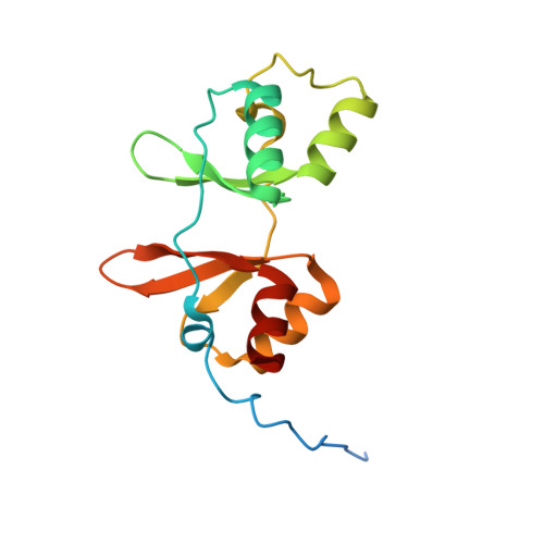
Cystathionine β-synthase (CBS) is a key enzyme in sulfur metabolism, and its inherited deficiency causes homocystinuria. Mammalian CBS is modulated by the binding of S-adenosyl-l-methionine (AdoMet) to its regulatory domain, which activates its catalytic domain. To investigate the underlying mechanism, we performed x-ray crystallography, mutagenesis, and mass spectrometry (MS) on human CBS. The 1.7 Å structure of a AdoMet-bound CBS regulatory domain shows one AdoMet molecule per monomer, at the interface between two constituent modules (CBS-1, CBS-2). AdoMet binding is accompanied by a reorientation between the two modules, relative to the AdoMet-free basal state, to form interactions with AdoMet via residues verified by mutagenesis to be important for AdoMet binding (Phe(443), Asp(444), Gln(445), and Asp(538)) and for AdoMet-driven inter-domain communication (Phe(443), Asp(538)). The observed structural change is further supported by ion mobility MS, showing that as-purified CBS exists in two conformational populations, which converged to one in the presence of AdoMet. We therefore propose that AdoMet-induced conformational change alters the interface and arrangement between the catalytic and regulatory domains within the CBS oligomer, thereby increasing the accessibility of the enzyme active site for catalysis.
- From the Structural Genomics Consortium, Nuffield Department of Clinical Medicine, University of Oxford, Oxford OX3 7DQ, United Kingdom.
Organizational Affiliation: