4URI

Crystal structure of chitinase-like agglutinin RobpsCRA from Robinia pseudoacacia


Experimental Data Snapshot

  • Method: X-RAY DIFFRACTION
  • Resolution: 1.85 Å
  • R-Value Free: 0.187 
  • R-Value Work: 0.148 
  • R-Value Observed: 0.150 

wwPDB Validation   3D Report Full Report


This is version 1.2 of the entry. See complete history


Literature

Structural Basis for Carbohydrate Binding Properties of a Plant Chitinase-Like Agglutinin with Conserved Catalytic Machinery.

Sulzenbacher, G.Roig-Zamboni, V.Peumans, W.J.Henrissat, B.Van Damme, E.J.Bourne, Y.

(2015) J Struct Biol 190: 115

  • DOI: https://doi.org/10.1016/j.jsb.2015.01.013
  • Primary Citation of Related Structures:  
    4URI

  • PubMed Abstract: 

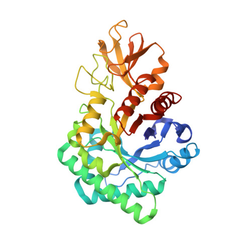
    A new chitinase-like agglutinin, RobpsCRA, related to family GH18 chitinases, has previously been identified in black locust (Robinia pseudoacacia) bark. The crystal structure of RobpsCRA at 1.85Å resolution reveals unusual molecular determinants responsible for the lack of its ancestral chitinase activity. Unlike other chitinase-like proteins, which lack chitinase catalytic residues, RobpsCRA has conserved its catalytic machinery. However, concerted rearrangements of loop regions coupled to non-conservative substitutions of aromatic residues central to the chitin-binding groove explain the lack of hydrolytic activity against chitin and the switch toward recognition of high-mannose type N-glycans. Identification of close homologs in flowering plants with conservation of sequence motifs associated to the structural adaptations seen in RobpsCRA defines an emerging class of agglutinins, as emphasized by a phylogenetic analysis, that are likely to share a similar carbohydrate binding specificity for high-mannose type N-glycans. This study illustrates the recent evolution and molecular adaptation of a versatile TIM-barrel scaffold within the ancestral GH18 family.


  • Organizational Affiliation

    Aix-Marseille University, AFMB UMR 7257, 13288 Marseille, France; CNRS, AFMB UMR 7257, 13288 Marseille, France.


Macromolecules
Find similar proteins by:  (by identity cutoff)  |  3D Structure
Entity ID: 1
MoleculeChains Sequence LengthOrganismDetailsImage
CHITINASE-RELATED AGGLUTININ
A, B
337Robinia pseudoacaciaMutation(s): 0 
UniProt
Find proteins for A1YZD2 (Robinia pseudoacacia)
Explore A1YZD2 
Go to UniProtKB:  A1YZD2
Entity Groups  
Sequence Clusters30% Identity50% Identity70% Identity90% Identity95% Identity100% Identity
UniProt GroupA1YZD2
Sequence Annotations
Expand
  • Reference Sequence
Small Molecules
Ligands 3 Unique
IDChains Name / Formula / InChI Key2D Diagram3D Interactions
MPD
Query on MPD

Download Ideal Coordinates CCD File 
C [auth A]
D [auth A]
E [auth A]
J [auth B]
K [auth B]
C [auth A],
D [auth A],
E [auth A],
J [auth B],
K [auth B],
L [auth B]
(4S)-2-METHYL-2,4-PENTANEDIOL
C6 H14 O2
SVTBMSDMJJWYQN-YFKPBYRVSA-N
CL
Query on CL

Download Ideal Coordinates CCD File 
I [auth A],
M [auth B]
CHLORIDE ION
Cl
VEXZGXHMUGYJMC-UHFFFAOYSA-M
NA
Query on NA

Download Ideal Coordinates CCD File 
F [auth A],
G [auth A],
H [auth A]
SODIUM ION
Na
FKNQFGJONOIPTF-UHFFFAOYSA-N
Experimental Data & Validation

Experimental Data

  • Method: X-RAY DIFFRACTION
  • Resolution: 1.85 Å
  • R-Value Free: 0.187 
  • R-Value Work: 0.148 
  • R-Value Observed: 0.150 
  • Space Group: P 1
Unit Cell:
Length ( Å )Angle ( ˚ )
a = 43.1α = 106.01
b = 53.221β = 94.12
c = 71.271γ = 107.59
Software Package:
Software NamePurpose
REFMACrefinement
MOSFLMdata reduction
SCALAdata scaling
PHASERphasing

Structure Validation

View Full Validation Report


Entry History 

Deposition Data

Revision History  (Full details and data files)

  • Version 1.0: 2015-03-11
    Type: Initial release
  • Version 1.1: 2015-05-13
    Changes: Database references
  • Version 1.2: 2024-01-10
    Changes: Data collection, Database references, Derived calculations, Other, Refinement description