4UMB | pdb_00004umb

Structural analysis of substrate-mimicking inhibitors in complex with Neisseria meningitidis 3-deoxy-D-arabino-heptulosonate 7-phosphate synthase - the importance of accommodating the active site water


Experimental Data Snapshot

  • Method: X-RAY DIFFRACTION
  • Resolution: 2.17 Å
  • R-Value Free: 
    0.227 (Depositor), 0.233 (DCC) 
  • R-Value Work: 
    0.201 (Depositor), 0.208 (DCC) 
  • R-Value Observed: 
    0.202 (Depositor) 

Starting Model: experimental
View more details

wwPDB Validation 3D Report Full Report

Validation slider image for 4UMB

Ligand Structure Quality Assessment 


This is version 1.3 of the entry. See complete history

Literature

Structural Analysis of Substrate-Mimicking Inhibitors in Complex with Neisseria Meningitidis 3-Deoxy-D-Arabino-Heptulosonate 7-Phosphate Synthase - the Importance of Accommodating the Active Site Water.

Heyes, L.C.Reichau, S.Cross, P.J.Jameson, G.B.Parker, E.J.

(2014) Bioorg Chem 57: 242

  • DOI: https://doi.org/10.1016/j.bioorg.2014.08.003
  • Primary Citation Related Structures: 
    4UMA, 4UMB, 4UMC

  • PubMed Abstract: 

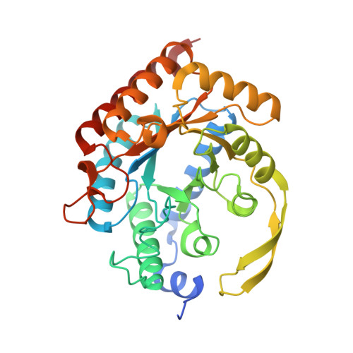
    3-Deoxy-d-arabino-heptulosonate 7-phosphate synthase (DAH7PS) catalyses the first committed step of the shikimate pathway, which produces the aromatic amino acids as well as many other aromatic metabolites. DAH7PS catalyses an aldol-like reaction between phosphoenolpyruvate and erythrose 4-phosphate. Three phosphoenolpyruvate mimics, (R)-phospholactate, (S)-phospholactate and vinyl phosphonate [(E)-2-methyl-3-phosphonoacrylate], were found to competitively inhibit DAH7PS from Neisseria meningitidis, which is the pathogen responsible for bacterial meningitis. The most potent inhibitor was the vinyl phosphonate with a Ki value of 3.9±0.4μM. We report for the first time crystal structures of these compounds bound in the active site of a DAH7PS enzyme which reveals that the inhibitors bind to the active site of the enzyme in binding modes that mimic those of the predicted oxocarbenium and tetrahedral intermediates of the enzyme-catalysed reaction. Furthermore, the inhibitors accommodate the binding of a key active site water molecule. Together, these observations provide strong evidence that this active site water participates directly in the DAH7PS reaction, enabling the facial selectivity of the enzyme-catalysed reaction sequence to be delineated.


  • Organizational Affiliation
    • Biomolecular Interaction Centre and Department of Chemistry, University of Canterbury, Christchurch, New Zealand.

Macromolecule Content 

  • Total Structure Weight: 156.13 kDa 
  • Atom Count: 10,942 
  • Modeled Residue Count: 1,334 
  • Deposited Residue Count: 1,404 
  • Unique protein chains: 1

Macromolecules

Find similar proteins by:|  3D Structure
Entity ID: 1
MoleculeChains  Sequence LengthOrganismDetailsImage
PHOSPHO-2-DEHYDRO-3-DEOXYHEPTONATE ALDOLASE
A, B, C, D
351Neisseria meningitidis MC58Mutation(s): 0 
EC: 2.5.1.54
UniProt
Find proteins for Q9K169 (Neisseria meningitidis serogroup B (strain ATCC BAA-335 / MC58))
Explore Q9K169 
Go to UniProtKB:  Q9K169
Entity Groups
Sequence Clusters30% Identity50% Identity70% Identity90% Identity95% Identity100% Identity
UniProt GroupQ9K169
Sequence Annotations
Expand
Reference Sequence

Small Molecules

Ligands 3 Unique
IDChains Name / Formula / InChI Key2D Diagram3D Interactions
0V5

Query on 0V5


Download:Ideal Coordinates CCD File
G [auth A],
I [auth B],
L [auth C],
O [auth D]
(2R)-2-(phosphonooxy)propanoic acid
C3 H7 O6 P
CSZRNWHGZPKNKY-UWTATZPHSA-N
PO4

Query on PO4


Download:Ideal Coordinates CCD File
F [auth A],
J [auth B],
M [auth C],
P [auth D]
PHOSPHATE ION
O4 P
NBIIXXVUZAFLBC-UHFFFAOYSA-K
MN

Query on MN


Download:Ideal Coordinates CCD File
E [auth A],
H [auth B],
K [auth C],
N [auth D]
MANGANESE (II) ION
Mn
WAEMQWOKJMHJLA-UHFFFAOYSA-N

Experimental Data & Validation

Experimental Data

  • Method: X-RAY DIFFRACTION
  • Resolution: 2.17 Å
  • R-Value Free:  0.227 (Depositor), 0.233 (DCC) 
  • R-Value Work:  0.201 (Depositor), 0.208 (DCC) 
  • R-Value Observed: 0.202 (Depositor) 
Space Group: P 21 21 21
Unit Cell:
Length ( Å )Angle ( ˚ )
a = 79.439α = 90
b = 133.733β = 90
c = 147.237γ = 90
Software Package:
Software NamePurpose
REFMACrefinement
iMOSFLMdata reduction

Structure Validation

View Full Validation Report


Ligand Structure Quality Assessment 


Entry History 

Deposition Data

Revision History  (Full details and data files)

  • Version 1.0: 2014-10-08
    Type: Initial release
  • Version 1.1: 2014-12-17
    Changes: Database references
  • Version 1.2: 2016-04-06
    Changes: Data collection
  • Version 1.3: 2024-01-10
    Changes: Data collection, Database references, Derived calculations, Other, Refinement description