


X-ray structure of the 4'-phosphopantetheinyl transferase PptT from Mycobacterium tuberculosis
Faille, A., Gavalda, S., Rottier, K., Mourey, L., Pedelacq, J.D.To be published.
Experimental Data Snapshot
Entity ID: 1 | |||||
|---|---|---|---|---|---|
| Molecule | Chains | Sequence Length | Organism | Details | Image |
| Phosphopantetheinyl transferase PptT | 259 | Mycobacterium tuberculosis H37Rv | Mutation(s): 0 Gene Names: pptT, Rv2794c, P425_02911, RVBD_2794c EC: 2.7.8.7 | 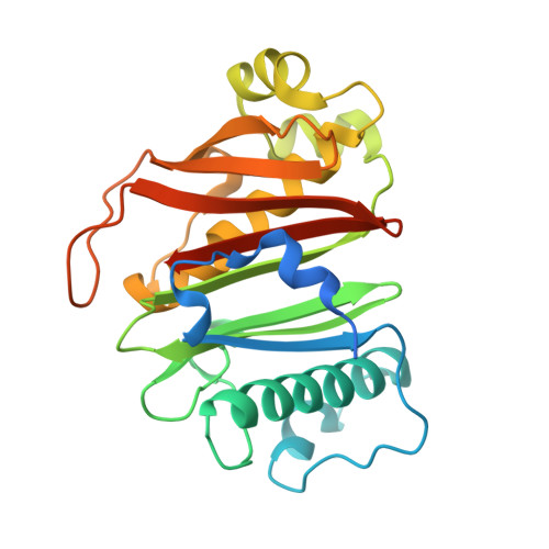 | |
UniProt | |||||
Entity Groups | |||||
| Sequence Clusters | 30% Identity50% Identity70% Identity90% Identity95% Identity100% Identity | ||||
| UniProt Group | O33336 | ||||
Sequence AnnotationsExpand | |||||
Reference Sequence | |||||
| Ligands 5 Unique | |||||
|---|---|---|---|---|---|
| ID | Chains | Name / Formula / InChI Key | 2D Diagram | 3D Interactions | |
| COA Download:Ideal Coordinates CCD File | B [auth A] | COENZYME A C21 H36 N7 O16 P3 S RGJOEKWQDUBAIZ-IBOSZNHHSA-N |  | ||
| PG4 Download:Ideal Coordinates CCD File | O [auth A] | TETRAETHYLENE GLYCOL C8 H18 O5 UWHCKJMYHZGTIT-UHFFFAOYSA-N |  | ||
| PO4 Download:Ideal Coordinates CCD File | N [auth A] | PHOSPHATE ION O4 P NBIIXXVUZAFLBC-UHFFFAOYSA-K |  | ||
| IMD Download:Ideal Coordinates CCD File | P [auth A] | IMIDAZOLE C3 H5 N2 RAXXELZNTBOGNW-UHFFFAOYSA-O |  | ||
| NA Download:Ideal Coordinates CCD File | C [auth A] D [auth A] E [auth A] F [auth A] G [auth A] | SODIUM ION Na FKNQFGJONOIPTF-UHFFFAOYSA-N |  | ||
| Length ( Å ) | Angle ( ˚ ) |
|---|---|
| a = 99.808 | α = 90 |
| b = 121.263 | β = 90 |
| c = 48.828 | γ = 90 |
| Software Name | Purpose |
|---|---|
| XDS | data reduction |
| SHELX | phasing |
| PDB_EXTRACT | data extraction |
| Coot | model building |
| REFMAC | refinement |
| XSCALE | data scaling |
| SHELXD | phasing |
| XSCALE | data reduction |


