4U5Y | pdb_00004u5y

Crystal Structure of the complex between the GNAT domain of S. lividans PAT and the acetyl-CoA synthetase C-terminal domain of S. enterica


Experimental Data Snapshot

  • Method: X-RAY DIFFRACTION
  • Resolution: 1.93 Å
  • R-Value Free: 
    0.239 (Depositor), 0.239 (DCC) 
  • R-Value Work: 
    0.187 (Depositor), 0.189 (DCC) 
  • R-Value Observed: 
    0.189 (Depositor) 

Starting Models: experimental
View more details

wwPDB Validation 3D Report Full Report

Validation slider image for 4U5Y

This is version 1.3 of the entry. See complete history

Literature

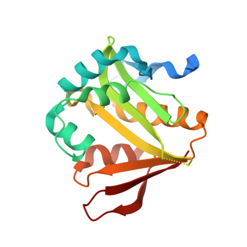
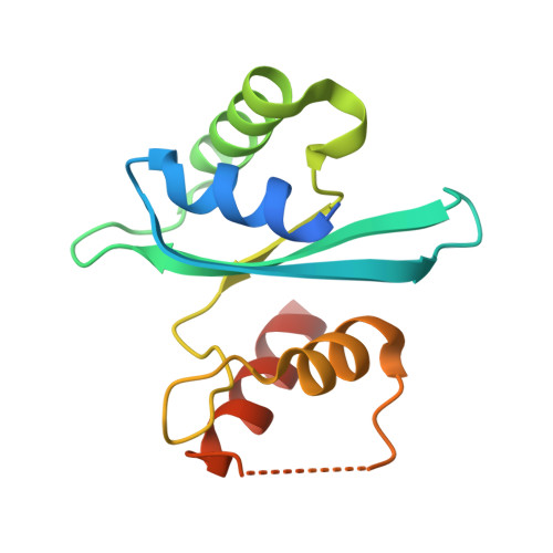
Insights into the specificity of lysine acetyltransferases.

Tucker, A.C.Taylor, K.C.Rank, K.C.Rayment, I.Escalante-Semerena, J.C.

(2014) J Biological Chem 289: 36249-36262

  • DOI: https://doi.org/10.1074/jbc.M114.613901
  • Primary Citation Related Structures: 
    4NXY, 4U5Y

  • PubMed Abstract: 

    Reversible lysine acetylation by protein acetyltransferases is a conserved regulatory mechanism that controls diverse cellular pathways. Gcn5-related N-acetyltransferases (GNATs), named after their founding member, are found in all domains of life. GNATs are known for their role as histone acetyltransferases, but non-histone bacterial protein acetytransferases have been identified. Only structures of GNAT complexes with short histone peptide substrates are available in databases. Given the biological importance of this modification and the abundance of lysine in polypeptides, how specificity is attained for larger protein substrates is central to understanding acetyl-lysine-regulated networks. Here we report the structure of a GNAT in complex with a globular protein substrate solved to 1.9 Å. GNAT binds the protein substrate with extensive surface interactions distinct from those reported for GNAT-peptide complexes. Our data reveal determinants needed for the recognition of a protein substrate and provide insight into the specificity of GNATs.


  • Organizational Affiliation
    • From the Department of Microbiology, University of Georgia, Athens, Georgia 30602 and.

Macromolecule Content 

  • Total Structure Weight: 36.4 kDa 
  • Atom Count: 2,396 
  • Modeled Residue Count: 280 
  • Deposited Residue Count: 329 
  • Unique protein chains: 2

Macromolecules

Find similar proteins by:|  3D Structure
Entity ID: 1
MoleculeChains  Sequence LengthOrganismDetailsImage
Acyl-CoA synthetase194Streptomyces lividans TK24Mutation(s): 1 
Gene Names: SSPG_01886
Find similar proteins by:|  3D Structure
Entity ID: 2
MoleculeChains  Sequence LengthOrganismDetailsImage
Acetyl-coenzyme A synthetaseB [auth D]135Salmonella enterica subsp. enterica serovar Paratyphi B str. ATCC 10719Mutation(s): 1 
Gene Names: acsSEEPB719_19728
EC: 6.2.1.1

Experimental Data & Validation

Experimental Data

  • Method: X-RAY DIFFRACTION
  • Resolution: 1.93 Å
  • R-Value Free:  0.239 (Depositor), 0.239 (DCC) 
  • R-Value Work:  0.187 (Depositor), 0.189 (DCC) 
  • R-Value Observed: 0.189 (Depositor) 
Space Group: C 2 2 21
Unit Cell:
Length ( Å )Angle ( ˚ )
a = 62.478α = 90
b = 66.524β = 90
c = 138.539γ = 90
Software Package:
Software NamePurpose
PHENIXrefinement

Structure Validation

View Full Validation Report


Entry History 

& Funding Information

Deposition Data


Funding OrganizationLocationGrant Number
USPHSUnited StatesGM062203
USPHSUnited StatesGM083987

Revision History  (Full details and data files)

  • Version 1.0: 2014-11-19
    Type: Initial release
  • Version 1.1: 2014-11-26
    Changes: Database references
  • Version 1.2: 2015-01-14
    Changes: Database references
  • Version 1.3: 2023-09-27
    Changes: Data collection, Database references, Derived calculations, Other, Refinement description, Source and taxonomy