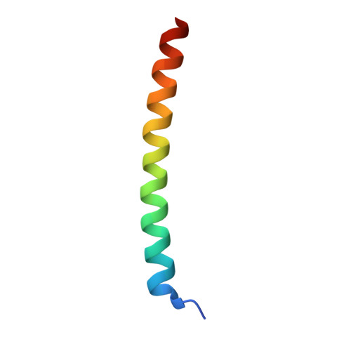
P6981, an arylstibonic acid, is a novel low nanomolar inhibitor of cAMP response element-binding protein binding to DNA.
Zhao, J., Stagno, J.R., Varticovski, L., Nimako, E., Rishi, V., McKinnon, K., Akee, R., Shoemaker, R.H., Ji, X., Vinson, C.(2012) Mol Pharmacol 82: 814-823
- PubMed: 22851716
- DOI: https://doi.org/10.1124/mol.112.080820
- Primary Citation of Related Structures:
4U5T - PubMed Abstract:
Several basic leucine zipper (B-ZIP) transcription factors have been implicated in cancer, substance abuse, and other pathological conditions. We previously identified arylstibonic acids that bind to B-ZIP proteins and inhibit their interaction with DNA. In this study, we used electrophoretic mobility shift assay to analyze 46 arylstibonic acids for their activity to disrupt the DNA binding of three B-ZIP [CCAAT/enhancer-binding protein α, cyclic AMP-response element-binding protein (CREB), and vitellogenin gene-binding protein (VBP)] and two basic helix-loop-helix leucine zipper (B-HLH-ZIP) [USF (upstream stimulating factor) and Mitf] proteins. Twenty-five arylstibonic acids showed activity at micromolar concentrations. The most active compound, P6981 [2-(3-stibonophenyl)malonic acid], had half-maximal inhibition at ~5 nM for CREB. Circular dichroism thermal denaturation studies indicated that P6981 binds both the B-ZIP domain and the leucine zipper. The crystal structure of an arylstibonic acid, NSC13778, bound to the VBP leucine zipper identified electrostatic interactions between both the stibonic and carboxylic acid groups of NSC13778 [(E)-3-(3-stibonophenyl)acrylic acid] and arginine side chains of VBP, which is also involved in interhelical salt bridges in the leucine zipper. P6981 induced GFP-B-ZIP chimeric proteins to partially localize to the cytoplasm, demonstrating that it is active in cells. P6981 inhibited the growth of a patient-derived clear cell sarcoma cell line whose oncogenic potential is driven by a chimeric protein EWS-ATF1 (Ewing's sarcoma protein-activating transcription factor 1), which contains the DNA binding domain of ATF1, a B-ZIP protein. NSC13778 inhibited the growth of xenografted clear cell sarcoma, and no toxicity was observed. These experiments suggest that antimony containing arylstibonic acids are promising leads for suppression of DNA binding activities of B-ZIP and B-HLH-ZIP transcription factors.
- Laboratory of Metabolism, National Cancer Institute, Bethesda, Maryland 20892, USA.
Organizational Affiliation:


