4U4S

Crystal structure of the GluA2 ligand-binding domain (S1S2J-L483Y-N754S) in complex with glutamate and BPAM25 at 1.90 A resolution.


Experimental Data Snapshot

  • Method: X-RAY DIFFRACTION
  • Resolution: 1.90 Å
  • R-Value Free: 0.208 
  • R-Value Work: 0.159 
  • R-Value Observed: 0.161 

wwPDB Validation   3D Report Full Report


Ligand Structure Quality Assessment 


This is version 1.4 of the entry. See complete history


Literature

Positive Allosteric Modulators of 2-Amino-3-(3-hydroxy-5-methylisoxazol-4-yl)propionic Acid Receptors Belonging to 4-Cyclopropyl-3,4-dihydro-2H-1,2,4-pyridothiadiazine Dioxides and Diversely Chloro-Substituted 4-Cyclopropyl-3,4-dihydro-2H-1,2,4-benzothiadiazine 1,1-Dioxides.

Francotte, P.Nrholm, A.B.Deva, T.Olsen, L.Frydenvang, K.Goffin, E.Fraikin, P.de Tullio, P.Challal, S.Thomas, J.Y.Iop, F.Louis, C.Botez-Pop, I.Lestage, P.Danober, L.Kastrup, J.S.Pirotte, B.

(2014) J Med Chem 57: 9539-9553

  • DOI: https://doi.org/10.1021/jm501268r
  • Primary Citation of Related Structures:  
    4U4S, 4U4X

  • PubMed Abstract: 

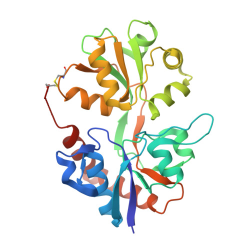
    Two 4-ethyl-substituted pyridothiadiazine dioxides belonging to α-amino-3-hydroxy-5-methyl-4-isoxazolepropionic acid (AMPA) receptor positive allosteric modulators were cocrystallized with the GluA2 ligand binding domain in order to decipher the impact of the position of the nitrogen atom on their binding mode at the AMPA receptors. The latter was found to be very similar to that of previously described benzothiadiazine-type AMPA receptor modulators. The affinity of the two compounds for the receptor was determined by isothermal titration calorimetry. Accordingly, the synthesis and biological evaluation of novel 4-cyclopropyl-substituted pyridothiadiazine dioxides was performed and completed with the synthesis of the corresponding chloro-substituted 4-cyclopropyl-3,4-dihydro-2H-benzothiadiazine 1,1-dioxides. The "8-aza" compound 32 was found to be the most potent pyridothiadiazine-type AMPA receptor potentiator in vitro, whereas the 7-chloro-substituted compound 36c emerged as the most promising benzothiadiazine dioxide. Due to proper drug-likeness and low in vivo acute toxicity in mice, 36c was chosen for a more complete preclinical evaluation. The compound was able to easily cross the blood-brain barrier. In an in vivo object recognition test with CD1 mice, oral administration of 36c was found to significantly improve cognition performance at doses as low as 1 mg/kg.


  • Organizational Affiliation

    Department of Medicinal Chemistry, Center for Interdisciplinary Research on Medicines (CIRM), University of Liege , Avenue de l'Hôpital, 1, B36, B-4000 Liège, Belgium.


Macromolecules
Find similar proteins by:  (by identity cutoff)  |  3D Structure
Entity ID: 1
MoleculeChains Sequence LengthOrganismDetailsImage
Glutamate receptor 2,Glutamate receptor 2
A, B
263Rattus norvegicusMutation(s): 2 
Gene Names: Gria2Glur2
UniProt
Find proteins for P19491 (Rattus norvegicus)
Explore P19491 
Go to UniProtKB:  P19491
Entity Groups  
Sequence Clusters30% Identity50% Identity70% Identity90% Identity95% Identity100% Identity
UniProt GroupP19491
Sequence Annotations
Expand
  • Reference Sequence
Small Molecules
Ligands 7 Unique
IDChains Name / Formula / InChI Key2D Diagram3D Interactions
3C1
Query on 3C1

Download Ideal Coordinates CCD File 
C [auth A],
N [auth B]
4-ethyl-3,4-dihydro-2H-pyrido[4,3-e][1,2,4]thiadiazine 1,1-dioxide
C8 H11 N3 O2 S
QAUSQKIKGJNUAS-UHFFFAOYSA-N
GLU
Query on GLU

Download Ideal Coordinates CCD File 
M [auth A],
X [auth B]
GLUTAMIC ACID
C5 H9 N O4
WHUUTDBJXJRKMK-VKHMYHEASA-N
PEG
Query on PEG

Download Ideal Coordinates CCD File 
R [auth B]DI(HYDROXYETHYL)ETHER
C4 H10 O3
MTHSVFCYNBDYFN-UHFFFAOYSA-N
SO4
Query on SO4

Download Ideal Coordinates CCD File 
D [auth A],
E [auth A],
O [auth B]
SULFATE ION
O4 S
QAOWNCQODCNURD-UHFFFAOYSA-L
GOL
Query on GOL

Download Ideal Coordinates CCD File 
F [auth A],
G [auth A],
P [auth B],
Q [auth B]
GLYCEROL
C3 H8 O3
PEDCQBHIVMGVHV-UHFFFAOYSA-N
ACT
Query on ACT

Download Ideal Coordinates CCD File 
H [auth A]
I [auth A]
J [auth A]
K [auth A]
S [auth B]
H [auth A],
I [auth A],
J [auth A],
K [auth A],
S [auth B],
T [auth B],
U [auth B],
V [auth B]
ACETATE ION
C2 H3 O2
QTBSBXVTEAMEQO-UHFFFAOYSA-M
CL
Query on CL

Download Ideal Coordinates CCD File 
L [auth A],
W [auth B]
CHLORIDE ION
Cl
VEXZGXHMUGYJMC-UHFFFAOYSA-M
Binding Affinity Annotations 
IDSourceBinding Affinity
3C1 Binding MOAD:  4U4S Kd: 1.20e+6 (nM) from 1 assay(s)
BindingDB:  4U4S Kd: 1.20e+6 (nM) from 1 assay(s)
Experimental Data & Validation

Experimental Data

  • Method: X-RAY DIFFRACTION
  • Resolution: 1.90 Å
  • R-Value Free: 0.208 
  • R-Value Work: 0.159 
  • R-Value Observed: 0.161 
  • Space Group: P 21 21 2
Unit Cell:
Length ( Å )Angle ( ˚ )
a = 97.83α = 90
b = 121.37β = 90
c = 47.33γ = 90
Software Package:
Software NamePurpose
SCALAdata scaling
PDB_EXTRACTdata extraction
PHENIXrefinement
PHASERphasing
Cootmodel building
XDSdata reduction

Structure Validation

View Full Validation Report


Ligand Structure Quality Assessment 


Entry History 

Deposition Data

Revision History  (Full details and data files)

  • Version 1.0: 2014-11-19
    Type: Initial release
  • Version 1.1: 2014-12-10
    Changes: Database references
  • Version 1.2: 2018-01-17
    Changes: Data collection
  • Version 1.3: 2018-06-13
    Changes: Data collection, Database references
  • Version 1.4: 2023-12-20
    Changes: Data collection, Database references, Refinement description