4U01 | pdb_00004u01

HCV NS3/4A serine protease in complex with 6570


Experimental Data Snapshot

  • Method: X-RAY DIFFRACTION
  • Resolution: 2.80 Å
  • R-Value Free: 
    0.290 (Depositor), 0.287 (DCC) 
  • R-Value Work: 
    0.233 (Depositor), 0.234 (DCC) 
  • R-Value Observed: 
    0.236 (Depositor) 

wwPDB Validation 3D Report Full Report

Validation slider image for 4U01

Ligand Structure Quality Assessment 


This is version 1.3 of the entry. See complete history

Literature

Discovery and structural diversity of the hepatitis C virus NS3/4A serine protease inhibitor series leading to clinical candidate IDX320.

Parsy, C.C.Alexandre, F.R.Bidau, V.Bonnaterre, F.Brandt, G.Caillet, C.Cappelle, S.Chaves, D.Convard, T.Derock, M.Gloux, D.Griffon, Y.Lallos, L.B.Leroy, F.Liuzzi, M.Loi, A.G.Moulat, L.Chiara, M.Rahali, H.Roques, V.Rosinovsky, E.Savin, S.Seifer, M.Standring, D.Surleraux, D.

(2015) Bioorg Med Chem Lett 25: 5427-5436

  • DOI: https://doi.org/10.1016/j.bmcl.2015.09.009
  • Primary Citation Related Structures: 
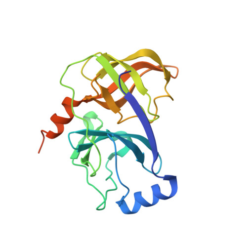
    4U01

  • PubMed Abstract: 

    Exploration of the P2 region by mimicking the proline motif found in BILN2061 resulted in the discovery of two series of potent HCV NS3/4A protease inhibitors. X-ray crystal structure of the ligand in contact with the NS3/4A protein and modulation of the quinoline heterocyclic region by structure based design and modeling allowed for the optimization of enzyme potency and cellular activity. This research led to the selection of clinical candidate IDX320 having good genotype coverage and pharmacokinetic properties in various species.


  • Organizational Affiliation
    • IDENIX, a wholly-owned subsidiary of Merck & Co, 1682 rue de la Valsière, Cap Gamma, BP 50001, 34189 Montpellier Cedex 4, France. Electronic address: christophe.parsy@merck.com.

Macromolecule Content 

  • Total Structure Weight: 215.98 kDa 
  • Atom Count: 13,360 
  • Modeled Residue Count: 1,755 
  • Deposited Residue Count: 1,971 
  • Unique protein chains: 2

Macromolecules

Find similar proteins by:|  3D Structure
Entity ID: 1
MoleculeChains  Sequence LengthOrganismDetailsImage
Non-structural 3 protease
A,
B,
C,
D,
E,
F,
G,
H,
I [auth J]
203Hepatitis C virus (isolate Con1)Mutation(s): 0 
UniProt
Find proteins for Q9WMX2 (Hepatitis C virus genotype 1b (isolate Con1))
Explore Q9WMX2 
Go to UniProtKB:  Q9WMX2
Entity Groups
Sequence Clusters30% Identity50% Identity70% Identity90% Identity95% Identity100% Identity
UniProt GroupQ9WMX2
Sequence Annotations
Expand
Reference Sequence
Find similar proteins by:  Sequence   |   3D Structure  
Entity ID: 2
MoleculeChains  Sequence LengthOrganismDetailsImage
NS4A protein16Orthohepacivirus hominisMutation(s): 0 
Gene Names: NS4A
UniProt
Find proteins for F0UY39 (Hepacivirus hominis)
Explore F0UY39 
Go to UniProtKB:  F0UY39
Entity Groups
Sequence Clusters30% Identity50% Identity70% Identity90% Identity95% Identity100% Identity
UniProt GroupF0UY39
Sequence Annotations
Expand
Reference Sequence

Small Molecules

Ligands 3 Unique
IDChains Name / Formula / InChI Key2D Diagram3D Interactions
39W

Query on 39W


Download:Ideal Coordinates CCD File
CA [auth E]
FA [auth F]
HA [auth G]
KA [auth H]
MA [auth J]
CA [auth E],
FA [auth F],
HA [auth G],
KA [auth H],
MA [auth J],
S [auth A],
U [auth B],
X [auth C],
Z [auth D]
(2S,3aS,10Z,11aS,12aR)-2-({8-fluoro-7-methoxy-2-[4-(propan-2-yl)-1,3-thiazol-2-yl]quinolin-4-yl}oxy)-5-methyl-N-[(1-methylcyclopropyl)sulfonyl]-4,14-dioxo-1,2,3,3a,4,5,6,7,8,9,11a,12,13,14-tetradecahydro-12aH-cyclopropa[m]pyrrolo[1,2-c][1,3,6]triazacyclotetradecine-12a-carboxamide
C37 H45 F N6 O7 S2
WCUAGYIETHEUQO-MULYSJGUSA-N
ZN

Query on ZN


Download:Ideal Coordinates CCD File
AA [auth D]
DA [auth E]
GA [auth F]
IA [auth G]
LA [auth H]
AA [auth D],
DA [auth E],
GA [auth F],
IA [auth G],
LA [auth H],
NA [auth J],
T [auth A],
V [auth B],
Y [auth C]
ZINC ION
Zn
PTFCDOFLOPIGGS-UHFFFAOYSA-N
CL

Query on CL


Download:Ideal Coordinates CCD File
BA [auth D],
EA [auth E],
JA [auth G],
W [auth B]
CHLORIDE ION
Cl
VEXZGXHMUGYJMC-UHFFFAOYSA-M

Experimental Data & Validation

Experimental Data

  • Method: X-RAY DIFFRACTION
  • Resolution: 2.80 Å
  • R-Value Free:  0.290 (Depositor), 0.287 (DCC) 
  • R-Value Work:  0.233 (Depositor), 0.234 (DCC) 
  • R-Value Observed: 0.236 (Depositor) 
Space Group: C 2 2 21
Unit Cell:
Length ( Å )Angle ( ˚ )
a = 150.034α = 90
b = 174.363β = 90
c = 133.01γ = 90
Software Package:
Software NamePurpose
REFMACrefinement
XDSdata reduction
XSCALEdata scaling

Structure Validation

View Full Validation Report


Ligand Structure Quality Assessment 


Entry History 

Revision History  (Full details and data files)

  • Version 1.0: 2015-07-29
    Type: Initial release
  • Version 1.1: 2015-10-14
    Changes: Database references
  • Version 1.2: 2015-11-11
    Changes: Database references
  • Version 1.3: 2024-05-08
    Changes: Data collection, Database references