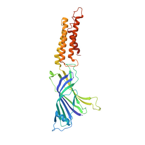
4TWF

X-ray structure of a pentameric ligand gated ion channel from Erwinia chrysanthemi (ELIC) in complex with bromomemantine


Experimental Data Snapshot

  • Method: X-RAY DIFFRACTION
  • Resolution: 3.90 Å
  • R-Value Free: 0.250 
  • R-Value Work: 0.202 
  • R-Value Observed: 0.204 

Starting Model: experimental
View more details

wwPDB Validation   3D Report Full Report


Ligand Structure Quality Assessment 


This is version 1.2 of the entry. See complete history


Literature

The Prokaryote Ligand-Gated Ion Channel ELIC Captured in a Pore Blocker-Bound Conformation by the Alzheimer's Disease Drug Memantine.

Ulens, C.Spurny, R.Thompson, A.J.Alqazzaz, M.Debaveye, S.Han, L.Price, K.Villalgordo, J.M.Tresadern, G.Lynch, J.W.Lummis, S.C.

(2014) Structure 22: 1399-1407

  • DOI: https://doi.org/10.1016/j.str.2014.07.013
  • Primary Citation of Related Structures:  
    4TWD, 4TWF, 4TWH

  • PubMed Abstract: 

    Pentameric ligand-gated ion channels (pLGIC) catalyze the selective transfer of ions across the cell membrane in response to a specific neurotransmitter. A variety of chemically diverse molecules, including the Alzheimer's drug memantine, block ion conduction at vertebrate pLGICs by plugging the channel pore. We show that memantine has similar potency in ELIC, a prokaryotic pLGIC, when it contains an F16'S pore mutation. X-ray crystal structures, using both memantine and its derivative, Br-memantine, reveal that the ligand is localized at the extracellular entryway of the channel pore, and the pore is in a more closed conformation than wild-type ELIC in both the presence and absence of memantine. However, using voltage clamp fluorometry we observe fluorescence changes in opposite directions during channel activation and pore block, revealing an additional conformational transition not apparent from the crystal structures. These results have important implications for drugs such as memantine, which block channel pores.


  • Organizational Affiliation

    Laboratory of Structural Neurobiology, KU Leuven, Leuven 3000, Belgium. Electronic address: chris.ulens@med.kuleuven.be.


Macromolecules
Find similar proteins by:  (by identity cutoff)  |  3D Structure
Entity ID: 1
MoleculeChains Sequence LengthOrganismDetailsImage
Cys-loop ligand-gated ion channel
A, B, C, D, E
A, B, C, D, E, F, G, H, I, J
307Dickeya chrysanthemiMutation(s): 1 
Membrane Entity: Yes 
UniProt
Find proteins for P0C7B7 (Dickeya chrysanthemi)
Explore P0C7B7 
Go to UniProtKB:  P0C7B7
Entity Groups  
Sequence Clusters30% Identity50% Identity70% Identity90% Identity95% Identity100% Identity
UniProt GroupP0C7B7
Sequence Annotations
Expand
  • Reference Sequence
Small Molecules
Ligands 1 Unique
IDChains Name / Formula / InChI Key2D Diagram3D Interactions
BR7
Query on BR7

Download Ideal Coordinates CCD File 
K [auth A]
L [auth A]
M [auth B]
N [auth C]
O [auth D]
K [auth A],
L [auth A],
M [auth B],
N [auth C],
O [auth D],
P [auth E],
Q [auth F],
R [auth F],
S [auth G],
T [auth H],
U [auth I],
V [auth J]
Bromomemantine
C12 H20 Br N
UKCUZUGMIJFTKF-IWDIQUIJSA-N
Experimental Data & Validation

Experimental Data

  • Method: X-RAY DIFFRACTION
  • Resolution: 3.90 Å
  • R-Value Free: 0.250 
  • R-Value Work: 0.202 
  • R-Value Observed: 0.204 
  • Space Group: P 1 21 1
Unit Cell:
Length ( Å )Angle ( ˚ )
a = 106.505α = 90
b = 266.092β = 107.7
c = 112.175γ = 90
Software Package:
Software NamePurpose
PHENIXrefinement
SCALAdata scaling
XDSdata reduction

Structure Validation

View Full Validation Report


Ligand Structure Quality Assessment 


Entry History 

Deposition Data

Revision History  (Full details and data files)

  • Version 1.0: 2014-09-24
    Type: Initial release
  • Version 1.1: 2014-10-22
    Changes: Database references
  • Version 1.2: 2023-12-20
    Changes: Data collection, Database references, Refinement description