4TUG | pdb_00004tug

Crystal structure of MjMre11-DNA2 complex


Experimental Data Snapshot

  • Method: X-RAY DIFFRACTION
  • Resolution: 3.55 Å
  • R-Value Free: 
    0.236 (Depositor), 0.237 (DCC) 
  • R-Value Work: 
    0.184 (Depositor), 0.189 (DCC) 
  • R-Value Observed: 
    0.187 (Depositor) 

wwPDB Validation 3D Report Full Report

Validation slider image for 4TUG

This is version 1.2 of the entry. See complete history

Literature

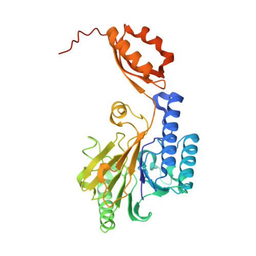
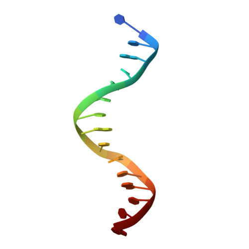
DNA end recognition by the Mre11 nuclease dimer: insights into resection and repair of damaged DNA.

Sung, S.Li, F.Park, Y.B.Kim, J.S.Kim, A.K.Song, O.K.Kim, J.Che, J.Lee, S.E.Cho, Y.

(2014) EMBO J 33: 2422-2435

  • DOI: https://doi.org/10.15252/embj.201488299
  • Primary Citation Related Structures: 
    4TUG, 4TUI

  • PubMed Abstract: 

    The Mre11-Rad50-Nbs1 (MRN) complex plays important roles in sensing DNA damage, as well as in resecting and tethering DNA ends, and thus participates in double-strand break repair. An earlier structure of Mre11 bound to a short duplex DNA molecule suggested that each Mre11 in a dimer recognizes one DNA duplex to bridge two DNA ends at a short distance. Here, we provide an alternative DNA recognition model based on the structures of Methanococcus jannaschii Mre11 (MjMre11) bound to longer DNA molecules, which may more accurately reflect a broken chromosome. An extended stretch of B-form DNA asymmetrically runs across the whole dimer, with each end of this DNA molecule being recognized by an individual Mre11 monomer. DNA binding induces rigid-body rotation of the Mre11 dimer, which could facilitate melting of the DNA end and its juxtaposition to an active site of Mre11. The identified Mre11 interface binding DNA duplex ends is structurally conserved and shown to functionally contribute to efficient resection, non-homologous end joining, and tolerance to DNA-damaging agents when other resection enzymes are absent. Together, the structural, biochemical, and genetic findings presented here offer new insights into how Mre11 recognizes damaged DNA and facilitates DNA repair.


  • Organizational Affiliation
    • Department of Life Sciences, Pohang University of Science and Technology, Pohang, South Korea.

Macromolecule Content 

  • Total Structure Weight: 247.36 kDa 
  • Atom Count: 16,104 
  • Modeled Residue Count: 1,891 
  • Deposited Residue Count: 2,051 
  • Unique protein chains: 1
  • Unique nucleic acid chains: 2

Macromolecules


Find similar proteins by:|  3D Structure
Entity ID: 1
MoleculeChains  Sequence LengthOrganismDetailsImage
DNA double-strand break repair protein Mre11337Methanocaldococcus jannaschii DSM 2661Mutation(s): 0 
Gene Names: mre11MJ1323
EC: 3.1
UniProt
Find proteins for Q58719 (Methanocaldococcus jannaschii (strain ATCC 43067 / DSM 2661 / JAL-1 / JCM 10045 / NBRC 100440))
Explore Q58719 
Go to UniProtKB:  Q58719
Entity Groups
Sequence Clusters30% Identity50% Identity70% Identity90% Identity95% Identity100% Identity
UniProt GroupQ58719
Sequence Annotations
Expand
Reference Sequence
Find similar nucleic acids by:  Sequence
Entity ID: 2
MoleculeChains LengthOrganismImage
DNA (5'-D(P*GP*CP*AP*CP*GP*TP*AP*GP*GP*AP*CP*AP*GP*C)-3')14synthetic construct
Sequence Annotations
Expand
Reference Sequence
Find similar nucleic acids by:  Sequence
Entity ID: 3
MoleculeChains LengthOrganismImage
DNA (5'-D(P*CP*TP*GP*TP*CP*CP*TP*AP*CP*GP*TP*GP*CP*CP*A)-3')15synthetic construct
Sequence Annotations
Expand
Reference Sequence

Small Molecules

Ligands 1 Unique
IDChains Name / Formula / InChI Key2D Diagram3D Interactions
MG

Query on MG


Download:Ideal Coordinates CCD File
I [auth C]
J [auth C]
K [auth A]
L [auth A]
M [auth B]
I [auth C],
J [auth C],
K [auth A],
L [auth A],
M [auth B],
N [auth B],
O [auth D],
P [auth D],
Q [auth E],
R [auth E],
S [auth F],
T [auth F]
MAGNESIUM ION
Mg
JLVVSXFLKOJNIY-UHFFFAOYSA-N

Experimental Data & Validation

Experimental Data

  • Method: X-RAY DIFFRACTION
  • Resolution: 3.55 Å
  • R-Value Free:  0.236 (Depositor), 0.237 (DCC) 
  • R-Value Work:  0.184 (Depositor), 0.189 (DCC) 
  • R-Value Observed: 0.187 (Depositor) 
Space Group: P 1 21 1
Unit Cell:
Length ( Å )Angle ( ˚ )
a = 91.53α = 90
b = 185.624β = 99.86
c = 106.212γ = 90
Software Package:
Software NamePurpose
PHENIXrefinement

Structure Validation

View Full Validation Report


Entry History 

Deposition Data

Revision History  (Full details and data files)

  • Version 1.0: 2014-10-15
    Type: Initial release
  • Version 1.1: 2014-10-29
    Changes: Database references
  • Version 1.2: 2023-12-27
    Changes: Data collection, Database references, Derived calculations, Other, Refinement description, Source and taxonomy