4TTR

Crystal structure of Legionella pneumophila dephospho-CoA kinase in complex with Bu2


Experimental Data Snapshot

  • Method: X-RAY DIFFRACTION
  • Resolution: 2.10 Å
  • R-Value Free: 0.228 
  • R-Value Work: 0.188 
  • R-Value Observed: 0.190 

wwPDB Validation   3D Report Full Report


This is version 1.3 of the entry. See complete history


Literature

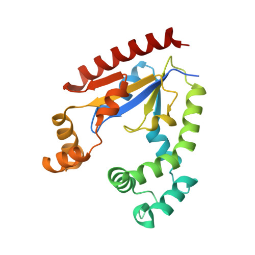
Crystal structure of Legionella pneumophila dephospho-CoA kinase reveals a non-canonical conformation of P-loop.

Gong, X.Chen, X.Yu, D.Zhang, N.Zhu, Z.Niu, L.Mao, Y.Ge, H.

(2014) J Struct Biol 188: 233-239

  • DOI: https://doi.org/10.1016/j.jsb.2014.10.008
  • Primary Citation of Related Structures:  
    4TTP, 4TTQ, 4TTR

  • PubMed Abstract: 
  • Dephospho-CoA kinase (DPCK; EC 2.7.1.24) catalyzes the final step in the coenzyme A biosynthetic pathway. DPCK transfers a phosphate group from ATP to the 3-hydroxyl group of the ribose of dephosphocoenzyme A (dCoA) to yield CoA and ADP. Upon the binding of ligands, large conformational changes is induced in DPCKs, as well as in many other kinases, to shield the bound ATP in their catalytic site from the futile hydrolysis by bulk water molecules ...

    Dephospho-CoA kinase (DPCK; EC 2.7.1.24) catalyzes the final step in the coenzyme A biosynthetic pathway. DPCK transfers a phosphate group from ATP to the 3-hydroxyl group of the ribose of dephosphocoenzyme A (dCoA) to yield CoA and ADP. Upon the binding of ligands, large conformational changes is induced in DPCKs, as well as in many other kinases, to shield the bound ATP in their catalytic site from the futile hydrolysis by bulk water molecules. To investigate the molecular mechanisms underlying the phosphoryl transfer during DPCK catalytic cycle, we determined the crystal structures of the Legionellapneumophila DPCK (LpDPCK) both in its apo-form and in complex with ATP. The structures reveal that LpDPCK comprises of three domains, the classical core domain, the CoA domain, and the LID domain, which are packed together to create a central cavity for substrate-binding and enzymatic catalysis. The binding of ATP induces large conformational changes, including a hinge-bending motion of the CoA binding domain and the "helix to loop" conformational change of the P-loop. Finally, modeling of a dCoA molecule to the enzyme provides insights into the catalytic mechanism of DPCK.


    Organizational Affiliation

    Institute of Health Sciences and School of Life Sciences, Anhui University, Hefei, Anhui 230601, China; Modern Experiment Technology Center, Anhui University, Hefei, Anhui 230601, China. Electronic address: gehonghua@gmail.com.


Macromolecules
Find similar proteins by:  (by identity cutoff)  |  3D Structure
Entity ID: 1
MoleculeChainsSequence LengthOrganismDetailsImage
Dephospho-CoA kinaseA215Legionella pneumophila subsp. pneumophila str. Philadelphia 1Mutation(s): 0 
Gene Names: coaElpg1467
EC: 2.7.1.24
UniProt
Find proteins for Q5ZVH3 (Legionella pneumophila subsp. pneumophila (strain Philadelphia 1 / ATCC 33152 / DSM 7513))
Explore Q5ZVH3 
Go to UniProtKB:  Q5ZVH3
Entity Groups  
Sequence Clusters30% Identity50% Identity70% Identity90% Identity95% Identity100% Identity
UniProt GroupQ5ZVH3
Protein Feature View
Expand
  • Reference Sequence
Small Molecules
Ligands 2 Unique
IDChainsName / Formula / InChI Key2D Diagram3D Interactions
PO4
Query on PO4

Download Ideal Coordinates CCD File 
C [auth A]PHOSPHATE ION
O4 P
NBIIXXVUZAFLBC-UHFFFAOYSA-K
 Ligand Interaction
BU2
Query on BU2

Download Ideal Coordinates CCD File 
B [auth A]1,3-BUTANEDIOL
C4 H10 O2
PUPZLCDOIYMWBV-BYPYZUCNSA-N
 Ligand Interaction
Experimental Data & Validation

Experimental Data

  • Method: X-RAY DIFFRACTION
  • Resolution: 2.10 Å
  • R-Value Free: 0.228 
  • R-Value Work: 0.188 
  • R-Value Observed: 0.190 
  • Space Group: P 21 21 2
Unit Cell:
Length ( Å )Angle ( ˚ )
a = 36.29α = 90
b = 82.2β = 90
c = 81.8γ = 90
Software Package:
Software NamePurpose
PHENIXrefinement

Structure Validation

View Full Validation Report


Entry History & Funding Information

Deposition Data

  • Released Date: 2014-12-10 
  • Deposition Author(s): Yu, D., Ge, H.

Funding OrganizationLocationGrant Number
National Natural Science Foundation of China (NSFC)China31270770

Revision History  (Full details and data files)

  • Version 1.0: 2014-12-10
    Type: Initial release
  • Version 1.1: 2014-12-24
    Changes: Database references
  • Version 1.2: 2015-10-07
    Changes: Experimental preparation
  • Version 1.3: 2020-01-08
    Changes: Author supporting evidence, Derived calculations