4TQQ

Photosynthetic Reaction Center from R. sphaeroides Analyzed at Room Temperature on an X-ray Transparent Microfluidic Chip


Experimental Data Snapshot

  • Method: X-RAY DIFFRACTION
  • Resolution: 2.50 Å
  • R-Value Free: 0.213 
  • R-Value Work: 0.162 
  • R-Value Observed: 0.165 

wwPDB Validation   3D Report Full Report


Ligand Structure Quality Assessment 


This is version 1.4 of the entry. See complete history


Literature

X-ray Transparent Microfluidic Chip for Mesophase-Based Crystallization of Membrane Proteins and On-Chip Structure Determination.

Khvostichenko, D.S.Schieferstein, J.M.Pawate, A.S.Laible, P.D.Kenis, P.J.

(2014) Cryst Growth Des 14: 4886-4890

  • DOI: https://doi.org/10.1021/cg5011488
  • Primary Citation of Related Structures:  
    4TQQ

  • PubMed Abstract: 

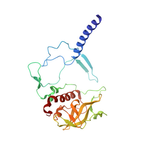
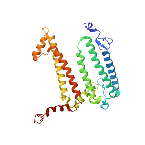
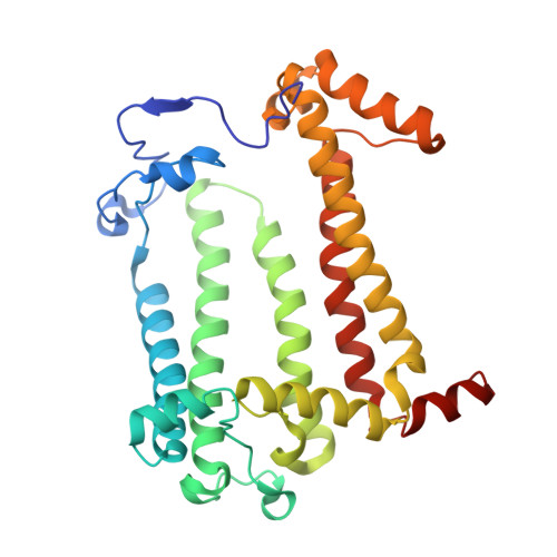
    Crystallization from lipidic mesophase matrices is a promising route to diffraction-quality crystals and structures of membrane proteins. The microfluidic approach reported here eliminates two bottlenecks of the standard mesophase-based crystallization protocols: (i) manual preparation of viscous mesophases and (ii) manual harvesting of often small and fragile protein crystals. In the approach reported here, protein-loaded mesophases are formulated in an X-ray transparent microfluidic chip using only 60 nL of the protein solution per crystallization trial. The X-ray transparency of the chip enables diffraction data collection from multiple crystals residing in microfluidic wells, eliminating the normally required manual harvesting and mounting of individual crystals. We validated our approach by on-chip crystallization of photosynthetic reaction center, a membrane protein from Rhodobacter sphaeroides , followed by solving its structure to a resolution of 2.5 Å using X-ray diffraction data collected on-chip under ambient conditions. A moderate conformational change in hydrophilic chains of the protein was observed when comparing the on-chip, room temperature structure with known structures for which data were acquired under cryogenic conditions.


  • Organizational Affiliation

    Department of Chemical & Biomolecular Engineering, University of Illinois at Urbana-Champaign , 600 South Mathews Avenue, Urbana, Illinois 61801, United States.


Macromolecules
Find similar proteins by:  (by identity cutoff)  |  3D Structure
Entity ID: 1
MoleculeChains Sequence LengthOrganismDetailsImage
Reaction center protein H chainA [auth H]240Cereibacter sphaeroidesMutation(s): 0 
Membrane Entity: Yes 
UniProt
Find proteins for P0C0Y7 (Cereibacter sphaeroides)
Explore P0C0Y7 
Go to UniProtKB:  P0C0Y7
Entity Groups  
Sequence Clusters30% Identity50% Identity70% Identity90% Identity95% Identity100% Identity
UniProt GroupP0C0Y7
Sequence Annotations
Expand
  • Reference Sequence
Find similar proteins by:  (by identity cutoff)  |  3D Structure
Entity ID: 2
MoleculeChains Sequence LengthOrganismDetailsImage
Reaction center protein L chainB [auth L]281Cereibacter sphaeroidesMutation(s): 0 
Membrane Entity: Yes 
UniProt
Find proteins for P0C0Y8 (Cereibacter sphaeroides)
Explore P0C0Y8 
Go to UniProtKB:  P0C0Y8
Entity Groups  
Sequence Clusters30% Identity50% Identity70% Identity90% Identity95% Identity100% Identity
UniProt GroupP0C0Y8
Sequence Annotations
Expand
  • Reference Sequence
Find similar proteins by:  (by identity cutoff)  |  3D Structure
Entity ID: 3
MoleculeChains Sequence LengthOrganismDetailsImage
Reaction center protein M chainC [auth M]302Cereibacter sphaeroidesMutation(s): 0 
Membrane Entity: Yes 
UniProt
Find proteins for P0C0Y9 (Cereibacter sphaeroides)
Explore P0C0Y9 
Go to UniProtKB:  P0C0Y9
Entity Groups  
Sequence Clusters30% Identity50% Identity70% Identity90% Identity95% Identity100% Identity
UniProt GroupP0C0Y9
Sequence Annotations
Expand
  • Reference Sequence
Small Molecules
Ligands 5 Unique
IDChains Name / Formula / InChI Key2D Diagram3D Interactions
BCL
Query on BCL

Download Ideal Coordinates CCD File 
E [auth L],
F [auth L],
J [auth M],
K [auth M]
BACTERIOCHLOROPHYLL A
C55 H74 Mg N4 O6
DSJXIQQMORJERS-AGGZHOMASA-M
BPH
Query on BPH

Download Ideal Coordinates CCD File 
D [auth L],
L [auth M]
BACTERIOPHEOPHYTIN A
C55 H76 N4 O6
KWOZSBGNAHVCKG-SZQBJALDSA-N
U10
Query on U10

Download Ideal Coordinates CCD File 
I [auth M]UBIQUINONE-10
C59 H90 O4
ACTIUHUUMQJHFO-UPTCCGCDSA-N
UQ1
Query on UQ1

Download Ideal Coordinates CCD File 
G [auth L]UBIQUINONE-1
C14 H18 O4
SOECUQMRSRVZQQ-UHFFFAOYSA-N
FE2
Query on FE2

Download Ideal Coordinates CCD File 
H [auth L]FE (II) ION
Fe
CWYNVVGOOAEACU-UHFFFAOYSA-N
Experimental Data & Validation

Experimental Data

  • Method: X-RAY DIFFRACTION
  • Resolution: 2.50 Å
  • R-Value Free: 0.213 
  • R-Value Work: 0.162 
  • R-Value Observed: 0.165 
  • Space Group: P 42 21 2
Unit Cell:
Length ( Å )Angle ( ˚ )
a = 102.484α = 90
b = 102.484β = 90
c = 239.939γ = 90
Software Package:
Software NamePurpose
HKL-2000data reduction
PHENIXrefinement
Cootmodel building
PDB_EXTRACTdata extraction

Structure Validation

View Full Validation Report


Ligand Structure Quality Assessment 


Entry History & Funding Information

Deposition Data


Funding OrganizationLocationGrant Number
National Institutes of Health/National Institute of General Medical Sciences (NIH/NIGMS)United StatesR01-GM086727

Revision History  (Full details and data files)

  • Version 1.0: 2014-07-16
    Type: Initial release
  • Version 1.1: 2014-10-22
    Changes: Database references
  • Version 1.2: 2017-09-13
    Changes: Author supporting evidence, Derived calculations, Other, Refinement description, Source and taxonomy
  • Version 1.3: 2019-12-25
    Changes: Author supporting evidence
  • Version 1.4: 2023-09-27
    Changes: Data collection, Database references, Derived calculations, Refinement description